When your doctor hands you an imaging requisition, it can feel like you have more questions than answers. Chest X-ray? Bone scan? DEXA? Barium swallow? The list of options can seem technical and intimidating, especially when you are already managing a health concern. This article breaks down the most common X-ray procedures, explains what actually happens during each one, and helps you understand why your healthcare provider might recommend a specific test. By the end, you will feel confident asking the right questions and knowing what to expect.
Table of Contents
- How X-rays work and what to expect
- The most common examples of X-ray procedures
- DEXA scans and bone density X-rays
- Contrast studies: Barium swallow and fluoroscopy
- Comparing X-ray procedures: Which is right for you?
- What most patients overlook when choosing an X-ray
- Take the next step with your X-ray care
- Frequently asked questions
Key Takeaways
| Point | Details |
|---|---|
| X-rays have many uses | They are critical for checking bones, lungs, teeth, and more. |
| Safety protocols matter | Modern imaging uses low radiation, and shielding is used thoughtfully when required. |
| DEXA scans check bone strength | These scans are ideal for osteoporosis screening, especially for higher-risk groups. |
| Contrast studies reveal more detail | Barium and fluoroscopy X-rays look at real-time movement, especially in the digestive tract. |
| Discuss choices with your provider | Your doctor helps select the best X-ray based on your health needs and safety. |
How X-rays work and what to expect
X-ray imaging is one of the oldest and most trusted tools in modern medicine, and the technology has come a long way. Understanding the basics helps you feel more prepared when you arrive at the clinic.
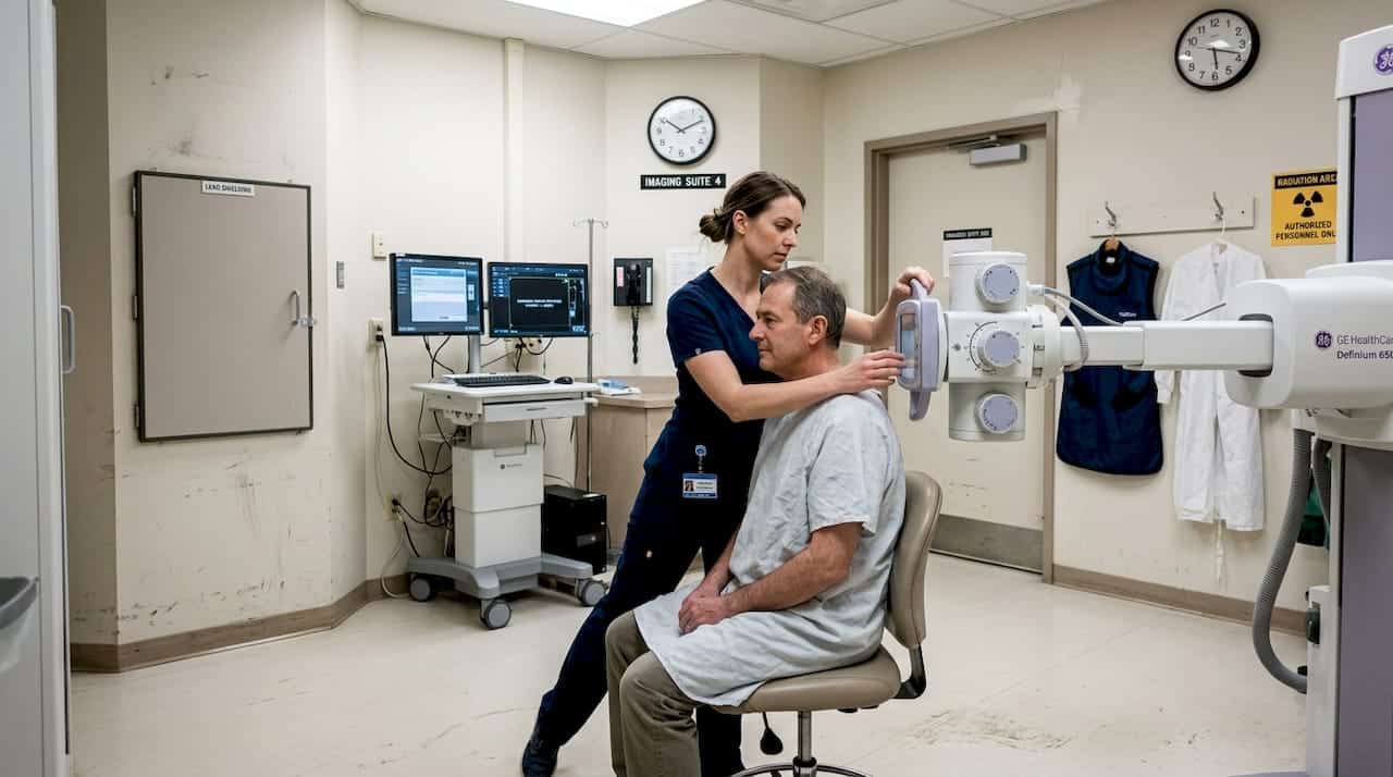
An X-ray machine emits ionising radiation that passes through your body. Dense tissues like bones absorb more radiation and appear white on the image, while soft tissues allow more radiation to pass through and appear grey or black. You are positioned by the technologist, asked to hold still or briefly hold your breath, and a digital detector captures the image in seconds. The whole process is quick and painless.
Here is what most patients experience during a standard X-ray appointment:
- Check-in and brief health history: You may be asked about pregnancy, recent surgeries, or implants.
- Changing or adjusting clothing: Metal objects, jewellery, and some clothing with fasteners are removed from the area being imaged.
- Positioning: The technologist will place you in the correct position, whether standing, sitting, or lying down.
- Image capture: You hold still for a second or two while the image is taken.
- Repeat views if needed: Two or three angles may be captured depending on the body part.
Modern digital X-ray systems are faster and more precise than older film technology, which means you can review your patient X-ray basics and generally expect fewer repeat exposures. Digital systems also allow faster turnaround on reports for your referring provider.
Safety note: Clinics follow the ALARA principle, which stands for “as low as reasonably achievable.” This means your imaging team uses the lowest radiation dose necessary to produce a clear, diagnostic-quality image. Always inform your technologist or ordering provider if you are pregnant or think you might be, as this affects positioning and shielding decisions. For a full overview of exposure levels and current X-ray safety information, it is always worth reviewing before your appointment.
Pro Tip: Wear comfortable, loose-fitting clothing to your appointment and leave metal jewellery at home. This saves time and avoids the need for a repeat exposure caused by artefacts in the image.
The most common examples of X-ray procedures
Understanding which type of X-ray is used for which body system makes it much easier to understand your referral. The types of X-rays your provider recommends will depend on your symptoms, your history, and the area of concern.
Here is a summary of the procedures you are most likely to encounter:
- Chest X-ray: Evaluates the lungs, heart size and shape, and surrounding structures. Commonly used for pneumonia, chronic obstructive pulmonary disease, heart enlargement, and pre-surgical screening.
- Bone X-ray: Focuses on a specific bone or joint. Used to identify fractures, monitor healing, assess arthritis severity, and track bone development in children.
- Dental X-ray: Taken in a dental office setting to detect cavities, root canal involvement, impacted teeth, and jaw structure changes.
- Abdominal X-ray: Examines the digestive organs, including the stomach, intestines, and kidneys. Helpful for identifying blockages, kidney stones, and signs of bowel obstruction.
- Spine X-ray: Looks at the vertebrae (bones of the spine) for scoliosis, disc space changes, arthritis, and alignment issues related to back pain.
Common X-ray tests span a wide range of clinical needs. The NHS confirms that these procedures cover a broad spectrum from lungs and heart to digestive organs, teeth, and the skeletal system, each tailored to the area and clinical question being answered.
| Procedure | Area imaged | Main uses | Common reasons for exam |
|---|---|---|---|
| Chest X-ray | Lungs, heart, ribs | Infection, heart changes, surgical prep | Cough, shortness of breath, chest pain |
| Bone X-ray | Limbs, joints, hands, feet | Fractures, arthritis, growth assessment | Injury, swelling, joint pain |
| Dental X-ray | Teeth, jaw, roots | Cavities, root damage, impaction | Routine dental checks, tooth pain |
| Abdominal X-ray | Stomach, bowel, kidneys | Stones, obstruction, organ size | Abdominal pain, nausea, bloating |
| Spine X-ray | Cervical, thoracic, lumbar spine | Scoliosis, disc spacing, arthritis | Back pain, postural concerns |
Each procedure has its own preparation and positioning requirements, so it helps to ask your clinic in advance what to expect for your specific exam.
DEXA scans and bone density X-rays
Beyond general X-rays, one special type plays a critical role in long-term bone health: the DEXA scan. DEXA stands for dual-energy X-ray absorptiometry, and it is specifically designed to measure how strong and dense your bones are.
A DEXA scan uses low-dose X-rays to assess bone mineral density and is a primary tool for osteoporosis screening. Osteoporosis is a condition where bones become weaker and more likely to fracture, often without obvious symptoms until a break occurs. Early detection through DEXA scanning can lead to earlier treatment and significantly better outcomes.
Here is what to expect, step by step:
- No special preparation is usually required. Avoid calcium supplements for 24 hours before your scan if advised by your provider.
- You remain fully clothed in most cases, though you will be asked to remove metal items.
- You lie flat on a padded table while the scanner arm passes slowly over your body. The exam is completely painless.
- The scanner measures two main sites: the lumbar spine and the hip, as these areas are most likely to experience fractures related to bone density loss.
- The scan takes 10 to 20 minutes and you can leave immediately afterward.
DEXA differs from a standard bone X-ray in an important way. A regular bone X-ray is primarily designed to spot structural changes like fractures or deformities, while a DEXA scan specifically quantifies bone mineral density and assigns a T-score that your provider uses to assess fracture risk. They serve different clinical purposes.
If you are looking for bone density testing in Southern Ontario, Valence Medical Imaging offers this service at multiple locations. If you are specifically searching for a bone density test in Brampton, or want to learn more about bone scan procedures before booking, detailed information is available online.
Pro Tip: DEXA scans are generally recommended for women over 65 and men over 70, but earlier screening is advised for anyone with a family history of osteoporosis, low body weight, or long-term steroid use. Speak with your family doctor about whether earlier screening is right for you.
Contrast studies: Barium swallow and fluoroscopy
Some X-ray procedures go beyond a single static image. Contrast studies use a special substance, most commonly barium, to make parts of your digestive tract visible on X-ray. Fluoroscopy is the real-time imaging technique used during these exams, allowing the radiologist to watch your body in motion.
A barium swallow is one of the most common contrast studies. You drink a thick, chalky liquid containing barium, which coats the lining of your oesophagus and stomach and shows up clearly on X-ray. The barium swallow procedure uses fluoroscopy to capture movement in real time, showing how you swallow, whether there are any structural abnormalities, and how well the oesophagus and stomach are functioning. If perforation of the digestive tract is suspected, water-soluble contrast is used instead of barium to avoid complications.
Here is what patients often want to know before a contrast study:
- The taste: Barium contrast has a chalky or slightly metallic taste. Some formulations are flavoured.
- The positioning: You may stand, sit, or lie on a tilting table that adjusts angle during the exam.
- Real-time images: The radiologist watches the images live on a monitor as you swallow, ensuring the right moments are captured.
- Duration: These exams typically take 15 to 30 minutes.
- After the exam: Drink plenty of water to help flush the barium from your system. Stools may appear white or pale for a day or two.
Safety reminder: Always tell your technologist or radiologist if you have a known allergy to contrast media, if you have difficulty swallowing, or if there is any chance you are pregnant. Patients with a high aspiration risk (where fluid may enter the airway) may need a modified procedure or a different type of contrast agent for safety.
For more information on fluoroscopy clinic services and what procedures are offered, you can browse options before your appointment to know exactly what to expect on the day.
Comparing X-ray procedures: Which is right for you?
Knowing the details of each procedure is useful, but a direct comparison can help you prepare for a conversation with your provider. The right exam depends on your symptoms, the body system being assessed, and the clinical question your doctor needs to answer.
Here is a quick comparison across key factors:
| Procedure | Purpose | Preparation needed | Comfort level | Radiation dose | Typical report time |
|---|---|---|---|---|---|
| Chest X-ray | Lungs, heart | Minimal | High | Very low | Same day to 48 hours |
| Bone X-ray | Fractures, arthritis | None | High | Very low | Same day to 48 hours |
| DEXA scan | Bone density | No supplements | Very high | Extremely low | 1 to 3 days |
| Abdominal X-ray | Digestive organs | Minimal | High | Low | Same day to 48 hours |
| Barium swallow | Oesophagus, stomach | Fasting required | Moderate | Low | 1 to 2 days |
| Spine X-ray | Vertebrae, discs | None | High | Low | Same day to 48 hours |
A standard chest X-ray, for example, has a dose area product of roughly 0.043 to 0.075 Gy·cm², which is well below the Danish diagnostic reference level of 0.3 Gy·cm². Modern protocols are continuously optimised to maintain image quality at the lowest possible dose, consistent with the ALARA principle.
Before your appointment, consider this checklist to discuss with your provider:
- What specific question is this X-ray intended to answer?
- Does the exam require any preparation, such as fasting or stopping certain supplements?
- Is there any alternative imaging (such as ultrasound or MRI) that might be more appropriate?
- Will I need follow-up imaging after this exam?
- How and when will I receive my results?
When comparing digital vs traditional X-rays, digital systems offer lower doses, faster results, and better image quality, making them the preferred standard at modern imaging clinics throughout Southern Ontario.
What most patients overlook when choosing an X-ray
After more than 35 years of serving patients across Southern Ontario, one pattern stands out clearly: the patients who get the most from their imaging experience are those who communicate openly with their care team rather than those who spend time researching radiation numbers alone.
There is a widespread belief that more lead shielding always means a safer X-ray. In reality, routine gonadal or lead shielding is no longer recommended for most exams. Modern imaging radiation risks are minimal because scatter radiation from today’s low-dose technology is extremely low. Placing shields over certain areas can actually obscure part of the image, which sometimes forces a repeat exposure, resulting in a higher total dose than if no shield had been used at all.
This does not mean shielding is never used. Pregnant patients should always inform their technologist, and abdominal shielding may be applied when it does not interfere with the clinical area being imaged. That distinction matters.
The most effective safety approach is not about demanding specific shielding protocols. It is about honest communication, telling your provider about your pregnancy status, prior surgeries, implants, and allergies, so the right decisions can be made for your specific situation. This is what what to know about X-rays consistently emphasises: informed patients get better outcomes.
Decision-making around imaging should focus on necessity, clinical appropriateness, and follow-up planning, not just radiation numbers. A well-chosen X-ray at the right time provides information that guides your care. An avoided or delayed X-ray because of unfounded concerns can mean a missed or late diagnosis.
Take the next step with your X-ray care
Valence Medical Imaging has supported patients across Southern Ontario for over 35 years, offering digital X-ray, DEXA bone density scanning, fluoroscopy, and more across seven clinic locations in Toronto, Scarborough, Brampton, Bramalea, Niagara Falls, and Whitby. With short wait times, fast report turnaround, and same-day availability for select services, getting the right imaging does not have to be complicated. Whether you are ready to book an X-ray appointment or want to explore your X-ray clinic options before making a decision, Valence makes it straightforward to move forward with your care. Your imaging team is here to guide you through every step.
Frequently asked questions
Are X-rays safe for children and pregnant patients?
Modern X-rays use low-dose technology, and shielding decisions for pregnant patients are made carefully based on the area being imaged and clinical need. Always inform your technologist of any pregnancy before the exam begins.
How should I prepare for an X-ray procedure?
Most X-rays require minimal preparation, primarily removing metal objects and informing your provider about pregnancy. Digital imaging has reduced the need for repeat exposures, but contrast studies like barium swallow typically require fasting beforehand.
What is a DEXA scan and who should get one?
A DEXA scan measures bone density using low-dose X-rays and is the standard test for osteoporosis screening. It is generally recommended for women over 65, men over 70, and younger adults with risk factors such as family history or long-term steroid use.
Can X-rays detect cancer or just bone problems?
X-rays can reveal tumours, unusual masses, and changes in bone or soft tissue that may indicate cancer, though some cancers require follow-up imaging such as CT, MRI, or biopsy for a definitive diagnosis.
Why might my doctor order a contrast X-ray like a barium swallow?
Contrast X-rays are used when standard imaging cannot clearly show movement or structural detail in areas like the digestive tract. Fluoroscopy with barium allows the radiologist to watch real-time function, which helps identify swallowing disorders, narrowing, or reflux that would not appear on a standard X-ray.