
Many people assume that all medical imaging involves radiation. In reality, diagnostic imaging covers a broad group of non-invasive techniques that use radiation, sound waves, magnetic fields, or contrast agents to create detailed pictures of the body’s interior. These tools help diagnose illness, assess severity, and guide treatment decisions. For patients across Southern Ontario, understanding what each type of imaging does and where to access it quickly can make a real difference in how confidently you navigate your healthcare. This article breaks down the most common types, what to expect, and how local clinics are making access easier than ever.
Table of Contents
- What is diagnostic imaging?
- Types of diagnostic imaging: X-ray, ultrasound, and mammography
- How do clinics in Southern Ontario deliver convenience and fast results?
- Nuances and things to keep in mind about imaging tests
- What most guides leave out about diagnostic imaging
- Book your imaging appointment for fast, clear answers
- Frequently asked questions
Key Takeaways
| Point | Details |
|---|---|
| Not all imaging uses radiation | Ultrasound relies on sound waves, making it safe for many, including pregnant patients. |
| Quick access is possible | Many clinics in Southern Ontario offer walk-in X-rays and rapid reporting. |
| Understand test limitations | Image quality and sensitivity vary by technique, so ask questions before your exam. |
| OHIP covers many exams | X-rays, ultrasounds, and mammograms are often available at no cost with a valid referral. |
What is diagnostic imaging?
Diagnostic imaging is a category of medical testing that allows healthcare providers to see inside your body without surgery. According to MedlinePlus, these are non-invasive techniques that use radiation, sound waves, magnetic fields, or contrast agents to create pictures of the body’s interior for diagnosing disorders, determining severity, and monitoring treatment.
That definition covers a wide range of tools. From the simple chest X-ray your family doctor orders to the detailed ultrasound used during pregnancy, diagnostic imaging is a cornerstone of modern medicine. It gives clinicians a way to see what is happening inside the body without guesswork.
Why does this matter for you? Because early detection changes outcomes. A mammogram that catches a small abnormality before symptoms appear, or an ultrasound that identifies a cyst before it causes complications, can significantly alter the course of treatment. Imaging is not just for emergencies. It is a routine part of preventive care, chronic disease management, and surgical planning.
Here are the most common reasons a healthcare provider might refer you for diagnostic imaging:
- Unexplained pain or swelling in any part of the body
- Suspected fractures, joint damage, or bone conditions
- Monitoring the progression of a known condition such as cancer or arthritis
- Evaluating organ function, including the liver, kidneys, or thyroid
- Screening for breast cancer or other conditions in asymptomatic patients
- Assessing vascular health, including blood flow and vein conditions
- Guiding procedures such as biopsies or drain placements
“Diagnostic imaging has transformed the way providers detect and manage disease. It reduces the need for exploratory surgery and allows for more targeted, effective treatment plans.”
It is also worth noting that not all imaging is the same. The type your provider orders depends on what they are looking for, which part of the body is involved, and whether radiation is a concern. Understanding your options helps you have a more informed conversation with your care team. You can also learn about OHIP coverage for mammograms if cost is a factor in your decision.
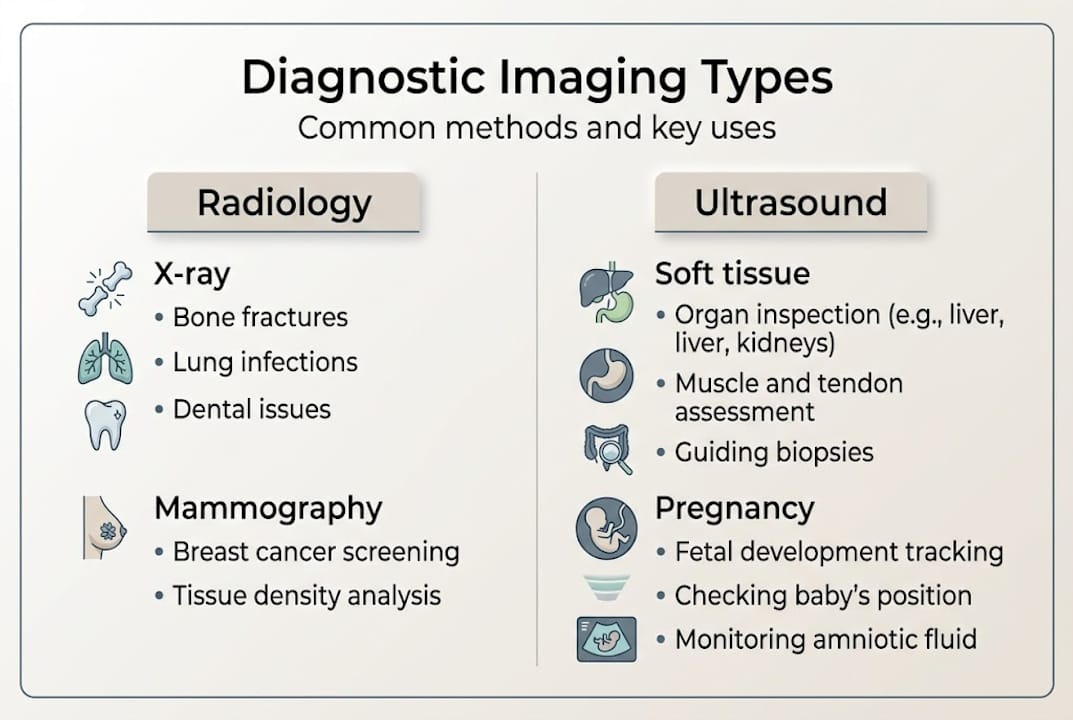
Types of diagnostic imaging: X-ray, ultrasound, and mammography
Now that we have defined diagnostic imaging, let’s look at the specific tools most commonly used, and what makes each one unique.
X-ray is the oldest and most widely used form of diagnostic imaging. It works by passing ionizing radiation through the body. Dense structures like bones absorb more radiation and appear white on the image, while soft tissues appear in shades of grey. X-rays are fast, widely available, and excellent for assessing fractures, lung conditions, and joint alignment. They are typically the first test ordered when a bone injury is suspected. However, because they use radiation, there are guidelines around how frequently they should be performed. You can read more about X-ray safety to understand what those guidelines mean for you.

Ultrasound works very differently. It uses high-frequency sound waves emitted by a transducer with piezoelectric crystals. These waves reflect off tissues based on differences in acoustic impedance, producing real-time two-dimensional images. Because ultrasound uses no ionizing radiation, it is considered very safe and is the preferred choice for imaging during pregnancy. It is also highly effective for soft tissue structures including the gallbladder, liver, kidneys, ovaries, and blood vessels. One limitation is that image quality can be affected by the angle of the transducer and by body composition, so operator skill matters considerably.
Mammography is a specialised form of X-ray designed specifically for breast tissue. It uses low-dose radiation to produce detailed images that can detect abnormalities too small to feel. Screening mammography is a key tool in early breast cancer detection, and Ontario’s Breast Screening Programme (OBSP) makes it accessible to eligible women. For patients who want the most detailed view of breast tissue, 3D mammography options are also available at select clinics.
Here is a quick comparison of the three main imaging types:
| Imaging type | Technology used | Radiation? | Best suited for | Speed |
|---|---|---|---|---|
| X-ray | Ionizing radiation | Yes (low dose) | Bones, lungs, fractures | Very fast |
| Ultrasound | Sound waves | No | Soft tissue, pregnancy, vascular | Moderate |
| Mammography | Low-dose X-ray | Yes (very low dose) | Breast tissue screening | Moderate |
Key considerations when choosing between these options include:
- Safety profile: Ultrasound is the only option with zero radiation exposure
- Sensitivity: Mammography is highly sensitive for breast tissue; ultrasound excels for soft organs
- Availability: X-ray is the most widely available and often walk-in friendly
- Follow-up needs: One test often leads to another for confirmation or detail
Pro Tip: If your provider orders an ultrasound and you are unsure why X-ray was not used instead, ask them directly. The choice of modality is deliberate and based on what tissue or structure needs to be assessed.
How do clinics in Southern Ontario deliver convenience and fast results?
A clear understanding of imaging types naturally leads to questions about where and how you can access them in Southern Ontario.
Access to diagnostic imaging in Southern Ontario has improved significantly in recent years. Walk-in X-ray is now available at many private clinics and some hospital-affiliated sites across the region, meaning you can often get imaged the same day you receive a referral. Ultrasound and mammography typically require an appointment, but booking windows at private clinics are generally much shorter than at hospital imaging departments.
OHIP covers most diagnostic imaging services when ordered by a physician or nurse practitioner. This includes X-ray, ultrasound, and screening mammography through the OBSP. For patients looking for OHIP mammogram locations in Brampton and surrounding areas, several approved sites offer both public and private booking options.
Here is what the typical patient journey looks like at a well-run Southern Ontario imaging clinic:
| Step | What happens | Typical time |
|---|---|---|
| Referral received | Provider sends requisition digitally or you bring a paper copy | Same day |
| Booking | Call or book online; same-day often available for X-ray | Minutes |
| Arrival | Check-in, brief intake, change if needed | 5-10 minutes |
| Imaging | Scan performed by registered technologist | 10-30 minutes |
| Report | Results sent to your provider, often within 24-48 hours | 1-2 business days |
What makes private clinics stand out is their focus on shorter waits and fast reports to providers, which is increasingly important as patients and physicians expect timely answers. Waiting weeks for a result creates anxiety and can delay treatment decisions. Clinics that prioritise turnaround time are directly improving patient outcomes.
When you arrive for your appointment, here is what you can generally expect:
- Bring your requisition form (paper or digital) and your health card
- Wear comfortable, loose clothing, particularly for abdominal or pelvic ultrasounds
- Avoid eating or drinking for several hours before certain abdominal ultrasounds
- Arrive a few minutes early to complete any intake paperwork
- Ask the technologist any questions you have before the scan begins
Southern Ontario’s network of imaging clinics continues to grow, with locations in cities including Toronto, Scarborough, Brampton, Bramalea, Niagara Falls, and Whitby. Whether you need a routine X-ray or a specialised scan, there is likely a clinic near you with short wait times and experienced staff.
Nuances and things to keep in mind about imaging tests
Even with convenience and rapid reporting, there are some factors every patient should be aware of before booking their scan.
Diagnostic imaging is a powerful tool, but it is not without limitations. Understanding these nuances helps you have realistic expectations and make more informed decisions alongside your healthcare provider.
Here are the key factors to keep in mind:
Image quality is task-dependent. The clarity of an image depends on the type of test, the equipment used, and the area being scanned. Factors like resolution, contrast, noise, and artifacts all affect what the radiologist can see. Modern AI-assisted reconstruction tools are improving noise reduction, but no test is perfect.
Radiation exposure is low but cumulative. For X-ray and mammography, radiation risks are low but do accumulate over a lifetime of imaging. This is why providers order only what is clinically necessary. If you have concerns, ask about radiation protection during X-ray, including the use of lead aprons.
Ultrasound is angle-dependent. Because sound waves reflect differently based on the angle of the transducer, some structures may be harder to visualise depending on body composition or the patient’s position. Doppler ultrasound, which measures motion and blood flow, has its own set of technical considerations.
Mammography involves compression and some discomfort. The breast must be compressed between two plates to produce a clear image. This can be uncomfortable, particularly for patients with sensitive breast tissue. The discomfort is brief and the compression is necessary for image quality.
False positives occur in approximately 10% of mammograms. Dense breast tissue can reduce sensitivity and increase the likelihood of a false positive result, which may lead to additional imaging or biopsy. This is not a failure of the test; it is a known trade-off in screening programmes.
“A false positive result can be stressful, but it is far better than a missed diagnosis. Follow-up testing is a routine part of the screening process, not a cause for alarm.”
Pro Tip: Before your appointment, write down any symptoms, previous imaging history, or medications you are taking. Sharing this information with the technologist helps ensure the right protocol is used and gives the radiologist important context when reading your images.
If you are uncertain about any aspect of your upcoming scan, do not hesitate to call the clinic ahead of time. A good imaging centre will take the time to answer your questions and help you feel prepared. You can also speak with your referring provider about mammogram OHIP coverage and whether any follow-up tests would be covered as well.
What most guides leave out about diagnostic imaging
Most articles about diagnostic imaging focus on what each test does and how it works. That information is useful, but it misses something important: what experienced patients do differently to get more value from their appointments.
The patients who navigate the system most effectively are not just passive recipients of care. They ask for copies of their results. They keep a personal health file with previous imaging reports and images. They ask their provider to explain what the radiologist found, not just whether the result was “normal” or “abnormal.” These habits create a clearer picture of health over time and help catch patterns that a single test might miss.
We also see patients who choose a clinic based purely on location or cost, without asking about report turnaround, radiologist credentials, or whether the clinic communicates results clearly to their family doctor. These details matter. A fast scan with a delayed or unclear report does not serve you well.
If you are considering screening mammography, the walk-in mammogram guide for Ontario patients is a practical resource that covers same-day access, eligibility, and what to bring. Being prepared before you arrive makes the entire experience smoother and more productive.
Book your imaging appointment for fast, clear answers
At Valence Medical Imaging, we have been serving Southern Ontario patients for over 35 years from seven clinic locations across Toronto, Scarborough, Brampton, Bramalea, Niagara Falls, and Whitby. We offer digital X-ray, ultrasound, mammography, OBSP screening, fluoroscopy, bone density imaging, and vein clinic ultrasound. Our team prioritises short wait times, fast report turnaround, and a patient-first experience at every visit. Whether you need a routine X-ray or are booking same-day ultrasound in Brampton, we make it straightforward. For patients interested in the latest screening technology, we also offer advanced 3D mammography. Book online or call your nearest location today.
Frequently asked questions
Is diagnostic imaging safe for everyone?
Most tests are very safe, but some such as X-rays and mammograms use low doses of radiation, and radiation risks are cumulative, so your provider will help you weigh any risks based on your personal health history.
Can I get an X-ray without an appointment in Southern Ontario?
Yes, many local clinics offer walk-in X-ray services with OHIP coverage, making it easy to get imaged quickly after receiving a referral from your provider.
Do all clinics in Southern Ontario offer fast report turnaround?
Turnaround varies by clinic, but some clinics advertise shorter waits and fast report delivery to your provider, so it is worth asking when you book.
Is ultrasound safe during pregnancy?
Yes, ultrasound uses only high-frequency sound waves with no ionizing radiation, and it is widely considered the safest imaging option for monitoring pregnancy.
Does OHIP cover mammograms in Ontario?
Yes, most screening mammograms are covered for eligible Ontario residents at OHIP-approved clinics, particularly through the Ontario Breast Screening Programme for women aged 50 to 74.
Recommended
- What Is Digital X-Ray? – Advancements in Imaging Technology – Valence Medical Imaging
- What Is the Difference Between an X-Ray and an MRI? – Choosing the Right Imaging Test – Valence Medical Imaging
- Do X-Rays Hurt? – What to Expect During an Imaging Procedure – Valence Medical Imaging
- Can I Walk in for an X-Ray? – Understanding Same-Day Imaging Services – Valence Medical Imaging