
Modern digital X-ray technology has transformed what patients experience when they walk into a diagnostic imaging clinic. Where older film-based systems required chemical processing and long waits, today’s digital detectors produce clear, detailed images almost instantly, with significantly less radiation exposure. If you’ve ever wondered what actually happens during a digital X-ray, why results seem to arrive so quickly, or whether the process is safe for you and your family, you’re not alone. This guide breaks down how digital X-ray works, what the different types mean for your care, and why choosing a clinic with advanced technology genuinely matters for your health outcomes.
Table of Contents
- What is a digital X-ray and how does it work?
- Comparing digital X-ray types: CR vs DR
- Safety and comfort: what does digital X-ray mean for you?
- Common questions: efficiency, results, and storage
- Our take: why digital X-ray is revolutionising patient care
- Next steps: find digital X-ray services near you
- Frequently asked questions
Key Takeaways
| Point | Details |
|---|---|
| Lower radiation | Digital X-ray cuts radiation exposure by up to 80% compared to traditional film. |
| Faster results | Electronic images are processed instantly, so you spend less time waiting. |
| Efficient workflow | Clinics can see more patients and deliver smoother, more comfortable experiences. |
| Two main methods | Computed Radiography and Direct Digital Radiography each have unique advantages for clinics and patients. |
What is a digital X-ray and how does it work?
Most people’s mental picture of an X-ray involves a technologist slipping a stiff film cassette under a table, taking the image, and then disappearing into a darkroom for several minutes. That process, while reliable for decades, has largely been replaced by digital technology that works faster, stores images electronically, and exposes patients to less radiation.
Digital X-ray uses electronic detectors instead of film to capture X-ray images, enabling instant processing, enhancement, and storage. Rather than waiting for chemical development, the detector converts X-ray energy into an electronic signal. That signal is processed by software within seconds, producing a high-resolution image on a monitor that the radiologist can review, adjust, and share immediately.
Here is what happens step by step during a typical digital X-ray:
- You are positioned by a registered technologist, either standing, seated, or lying down, depending on the body part being examined.
- The X-ray machine directs a focused beam of radiation through the targeted area of your body.
- The electronic detector on the other side captures the energy that passes through, creating a digital signal.
- Software converts that signal into a detailed greyscale image within seconds.
- The image is reviewed on screen, and the radiologist can adjust brightness, contrast, and magnification without retaking the X-ray.
- The final image and report are stored securely in the clinic’s system and shared with your referring physician.
One of the most meaningful improvements for patients is the ability to enhance images after they are taken. If a specific area needs to be seen more clearly, the radiologist can adjust the image digitally rather than asking you to return for another exposure. This reduces both your time in the clinic and your overall radiation dose.
“Digital imaging has fundamentally changed the speed and quality of diagnostic radiology. Patients benefit from faster answers and clinicians benefit from images they can manipulate and share instantly.”
Pro Tip: When you book your appointment, ask whether the clinic uses direct digital radiography (DR) rather than older computed radiography (CR) equipment. DR produces results even faster and typically involves less radiation.
The digital X-ray benefits extend well beyond speed. Electronic storage means your images are never lost, never degrade over time, and can be retrieved instantly for follow-up appointments or specialist consultations. For busy clinics across Southern Ontario, this efficiency means shorter wait times and a smoother experience from check-in to results.
Comparing digital X-ray types: CR vs DR
Understanding that there are different flavours of digital X-ray, it’s helpful to see how each method stacks up for patient care and clinic workflow.
There are two main methodologies: Computed Radiography (CR) uses photostimulable phosphor plates scanned by laser (indirect); Direct Digital Radiography (DR) uses flat-panel detectors converting X-rays directly to electrical signals. Both are far superior to traditional film, but they differ in meaningful ways that affect your experience as a patient.
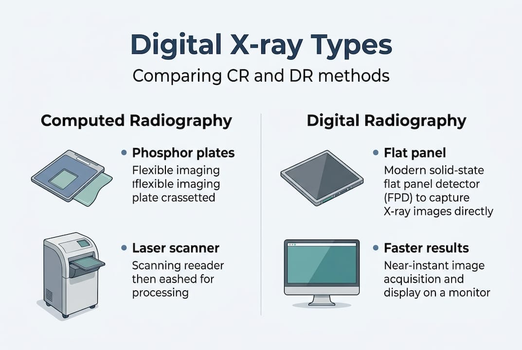
Computed Radiography (CR) was the first widely adopted form of digital X-ray. It replaced film with a reusable phosphor plate that stores the X-ray image temporarily. After exposure, the plate is fed into a separate reader that scans it with a laser and converts it to a digital file. This process typically takes between 30 and 90 seconds per image. CR is compatible with older X-ray equipment, which made it a cost-effective transition for many clinics moving away from film.
Direct Digital Radiography (DR) is the newer and more advanced standard. Instead of a phosphor plate, DR uses a flat-panel detector that converts X-ray energy directly into a digital signal. Images appear on the monitor in as little as 5 to 10 seconds. There is no separate scanning step, no plate to handle, and no delay between exposure and image review.
| Feature | Traditional film | CR | DR |
|---|---|---|---|
| Image processing time | 5 to 10 minutes | 30 to 90 seconds | 5 to 10 seconds |
| Radiation dose | Baseline | Reduced | Lowest |
| Image enhancement | Not possible | Limited | Full digital adjustment |
| Storage | Physical film | Digital file | Digital file |
| Equipment cost | Low | Moderate | Higher |
| Patient throughput | Slowest | Moderate | Fastest |
Key advantages of DR over CR:
- Faster image acquisition means less time on the table for patients with pain or mobility challenges.
- Superior image quality with higher resolution and greater detail in soft tissue and bone.
- Lower radiation dose per exposure, often 50 to 80% less than traditional film.
- Fewer repeat exposures needed because images can be adjusted digitally.
- Streamlined workflow allows technologists to see and confirm image quality before the patient leaves the room.
Statistic spotlight: DR systems offer a radiation dose reduction of 50 to 80% compared to traditional film X-ray, making them the preferred choice for clinics prioritising patient safety.
It is worth noting that some clinics in Southern Ontario still operate CR systems, particularly in older facilities or those that have not yet upgraded their equipment. If radiation minimisation and speed are priorities for you, asking specifically about DR technology before booking is a practical step. This is especially relevant for patients who require frequent imaging due to ongoing health conditions, where cumulative dose management becomes an important consideration. Understanding the difference between these two systems also helps explain why some clinics can offer faster turnaround on reports and why the imaging experience varies from one facility to another. Advances in 3D mammography technology follow a similar trajectory, where newer detector technology directly improves both safety and diagnostic accuracy.
Safety and comfort: what does digital X-ray mean for you?
With the technical comparisons in mind, let’s drill into what digital X-ray technology actually means for your safety and experience as a patient.
Digital X-rays reduce radiation dose by 50 to 80% compared to traditional film due to higher efficiency in how detectors capture X-ray energy, a measure known as Detective Quantum Efficiency (DQE). In practical terms, this means you receive a meaningful diagnostic image while absorbing far less radiation than patients did a generation ago. For most routine imaging, the dose from a digital X-ray is comparable to the natural background radiation you receive from the environment over a few hours to a few days.
Here is how digital X-ray improves the patient experience from start to finish:
- Shorter appointments. Because images are ready in seconds, your time in the imaging room is reduced. There is no waiting for film to develop before the technologist can confirm a clear image was captured.
- Fewer repeat exposures. Digital images can be brightened, darkened, or magnified after the fact. If a shadow or unclear area needs closer inspection, the radiologist adjusts the existing image rather than requesting a new exposure.
- Less repositioning. Faster image confirmation means the technologist can quickly verify positioning is correct before you move. This is particularly helpful for patients with joint pain, arthritis, or limited mobility.
- Comfortable for children. Paediatric patients often struggle to remain still for extended periods. The speed of DR imaging reduces the chance of motion blur, which means fewer retakes and a less stressful experience for young patients and their parents.
- Accessible for complex needs. Patients who use wheelchairs or have difficulty with certain positions benefit from the speed and flexibility of digital systems, which accommodate a wider range of positioning approaches.
“Reducing the time a patient spends in an uncomfortable position, combined with lower radiation exposure, represents a genuine improvement in the quality of care, not just the quality of the image.”
Consider a common scenario: you visit a clinic in Brampton or Whitby for a chest X-ray after your family doctor suspects a respiratory issue. With DR technology, you are in and out of the imaging room in under ten minutes. Your physician receives a digital report within hours, sometimes the same day. There is no waiting days for film to be processed, mailed, or retrieved from storage.
Pro Tip: If you are concerned about radiation, ask your technologist about the specific dose for your examination. Reputable clinics are transparent about this information and can put the numbers in context relative to everyday radiation exposure.
For patients who require X-ray safety information before their appointment, understanding DQE and dose reduction can make a real difference in how comfortable and confident you feel walking into the clinic.
Common questions: efficiency, results, and storage
Having looked at safety and comfort, let’s answer the practical questions patients ask most about their imaging experience and results.
How quickly are results available? Digital X-ray uses electronic storage for instant retrieval and sharing, supporting quicker reports and easier follow-ups. In a modern clinic using DR technology, the radiologist can begin reviewing your image within minutes of it being taken. Your referring physician typically receives a written report within hours, and in urgent cases, results can be communicated even faster through secure digital channels.
Can I access my own images? Many clinics now offer patient portals or provide images on a disc or secure digital link upon request. This is particularly useful if you are seeing multiple specialists or travelling for care. Having your images readily available avoids delays caused by tracking down physical films.
How are images stored? Digital X-ray files are stored in a system called a PACS (Picture Archiving and Communication System). This is a secure, searchable database that allows authorised healthcare providers to retrieve your images instantly, regardless of when the original study was performed. Images stored digitally do not degrade over time, unlike physical film, which can fade, crack, or be lost.
The table below compares efficiency across the three imaging approaches:
| Efficiency measure | Traditional film | CR | DR |
|---|---|---|---|
| Image ready for review | 5 to 10 minutes | 30 to 90 seconds | 5 to 10 seconds |
| Report turnaround | Hours to days | Hours | Minutes to hours |
| Image sharing | Physical delivery | Electronic | Instant electronic |
| Storage reliability | Degrades over time | Stable digital | Stable digital |
| Multi-specialist access | Difficult | Possible | Seamless |
Key practical benefits for patients:
- Same-day imaging is feasible and commonly offered at clinics with DR technology.
- Images can be shared instantly between your family doctor, specialist, and imaging centre without physical transport.
- Secure digital X-ray storage means your records are accessible for years without risk of physical deterioration.
- Booking a walk-in X-ray service is a realistic option at many Southern Ontario clinics, reducing the need to wait days for a scheduled appointment.
The efficiency of digital workflows also benefits patients indirectly. When clinics can process and share images faster, radiologists spend less time on administrative tasks and more time on careful image interpretation. That translates to more accurate, timely diagnoses for everyone in the system.
Our take: why digital X-ray is revolutionising patient care
We have been providing diagnostic imaging services to Southern Ontario patients for over 35 years, and the shift to digital X-ray is one of the most significant improvements we have seen in that time. Not because the technology is impressive for its own sake, but because it directly changes what patients experience and how quickly they get answers.
DR is preferred over CR for modern clinics due to speed and dose reduction, enhancing patient throughput and safety. What this means in practice is that more patients can be seen in a day, each with less radiation exposure and a faster path to diagnosis. For a family-owned clinic serving communities across Toronto, Scarborough, Brampton, Bramalea, Niagara Falls, and Whitby, that efficiency matters enormously.
Our honest advice: when you are choosing the best X-ray clinic in Southern Ontario, ask whether they use DR technology. It is not a minor detail. It affects your radiation dose, your wait time, and the speed at which your physician receives your results. Patients deserve to know the difference and to make informed choices about where they receive their care.
Next steps: find digital X-ray services near you
Armed with this knowledge, you are ready to choose a clinic that prioritises both your time and safety. At Valence Medical Imaging, we use advanced DR technology across our seven Southern Ontario locations, offering patients faster imaging, lower radiation exposure, and same-day availability for select services. Whether you need a routine chest X-ray or imaging for a specific concern, our team is committed to making the process as efficient and comfortable as possible.
To learn more about digital X-rays and what to expect at your appointment, visit our website for detailed patient resources. If you are ready to book, we offer flexible scheduling and the option to arrange a walk-in X-ray appointment at many of our locations. Our goal is to get you the answers you need, quickly and safely.
Frequently asked questions
How much radiation do I get from a digital X-ray?
Digital X-rays reduce radiation dose by 50 to 80% compared to film, thanks to advanced detector technology that captures images more efficiently with less exposure.
How quickly will I get my results from a digital X-ray?
Most digital X-ray images are available to your doctor in seconds to minutes after the exposure, because digital detectors enable instant processing and electronic delivery without any film development step.
Are digital X-rays safe for children?
Yes, digital X-rays are considered very safe for patients of all ages, and DR systems are preferred in modern clinics specifically because they deliver the lowest possible radiation dose while maintaining excellent image quality.
Is a digital X-ray appointment longer than a film X-ray?
Digital X-ray appointments are typically shorter than film-based ones, since CR processes images in 30 to 90 seconds and DR delivers results in as little as 5 to 10 seconds, significantly reducing your time in the imaging room.
Recommended
- What Is Digital X-Ray? – Advancements in Imaging Technology – Valence Medical Imaging
- Are X-Rays Safe? Understanding Radiation Exposure in Modern Imaging – Valence Medical Imaging
- How Are X-Rays Stored? – Digital vs. Film Imaging Explained – Valence Medical Imaging
- Can I Walk in for an X-Ray? – Understanding Same-Day Imaging Services – Valence Medical Imaging