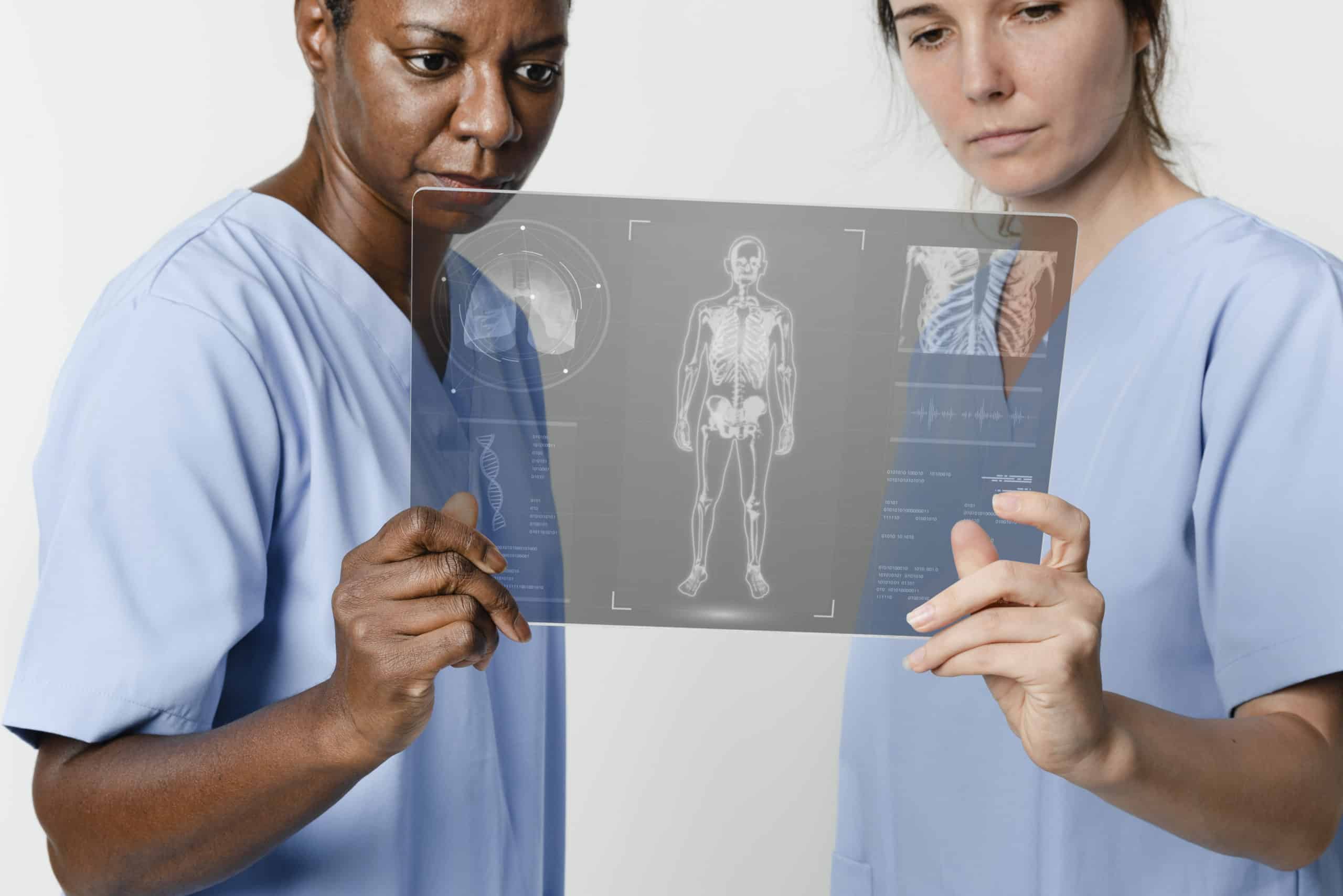
Doctors often rely on advanced diagnostic tools for better diagnosis and treatment planning. It is important to check out the X-rays before proceeding with any treatment. This not only helps in confirming the preliminary diagnosis but is also beneficial in identifying if there are any other related complications. X-ray imaging uses X-ray photons and creates images based on the density of the bones. They are popularly used for identifying fractures, cracks, bone density loss, etc. X-rays are mainly used for the diagnosis of bone-related medical disorders. They are comparatively inexpensive and less invasive methods of investigation that offer the best results.
Types of X-rays
There are mainly two types of X-rays that are popularly used: digital x-rays and traditional x-rays. Traditional X-rays have been in practice since the 1900s. They are an effective way of diagnosis. In this method, the patient is exposed in front of an X-ray machine. The position indicating the device that is the PID is targeted directly on the patient, around the target site. Once focused, a film is placed against the target organ, and an image is captured. This helps in clearly developing the images of the internal structure that help create an image.
In traditional X-rays, the image is developed on a film with the help of dyes and developer fluid. Many mishaps began to happen during the film development process, such as under-development or over-development of the film. This created a major concern in image diagnosis. To eliminate this process, the advanced technology of computers was incorporated into the field of X-rays. This gave rise to the creation of digital X-rays. Digital X-rays are comparatively more accurate and less expensive for patients. They also help provide faster images, thus, helping in faster diagnosis.
During the projection of a traditional X-ray, the patient is exposed to many harmful radiations that can negatively affect the health of the patient. Even though the amount of radiation exposure to the patient is very less, the doctor often takes advanced precautions. Some common preventive methods include wearing lead aprons, a thyroid collar, etc.
Pregnant ladies are also not allowed X-rays, especially in the first and third trimesters. This is because the X-rays hamper the process of organogenesis and can impact the growth of the fetus. The doctors also take special measures to ensure their protection. One of the measures taken is incorporating a glass window. The doctor often stands beyond it to avoid extensive radiation.
With the rise in digital X-rays, the side effects of X-rays and harmful exposures are reduced drastically. Almost 80% of the harmful radiation is reduced with digital X-rays. Thus, they are comparatively much safer and have almost no harmful effects due to repeated use. At the same time, since the side effects are reduced drastically, the doctors can easily take multiple X-rays without worrying about the adverse effects on the patients.
Traditional X-rays take a long time to develop. Moreover, the clarity of the image and the quality of the image greatly depend on the quality of the developer fluid used. The X-Ray film cannot be exposed to light before or after projection till the development process is completed. They must be developed in a developer fluid and rinsed properly to get the right image quality. If the developer fluid is contaminated or mixed, it hampers the image quality. The image is completely lost if the film is exposed to light before development. However, all these problems are completely omitted during the digital X Rays.
The cost involved in the development process includes installing a dark room. This is a four-by-four room with pitch-black walls. It is made in such a way that light can’t enter, making it easier to develop the X-ray film. Furthermore, the tanks have to be set up, where the fluid has to be stored. The developer fluid has to be replaced and added again, as per the use. On the other hand, only a machine has to be set up for digital X-rays. Thus, the cost has been reduced drastically.
For traditional X Rays, the patient, as well as the doctor, has to maintain many records. Since it is not possible to duplicate the image film, it is important to store and secure it in a safe place. If the film is misplaced, it is impossible to get it back. On the other hand, a digital X-Ray can easily be duplicated, shared and stored. It is as simple as saving an image on your phone. The image quality can also be edited and altered to get the right image effects. The doctor can also easily enlarge the image to the desired size.
Conclusion
Traditional X-rays were a great option in the past when there was a limit on the number of resources. However, today, when there is an option for digital X-rays, doctors must consider shifting to it, as it is a more reliable and convenient X-ray imaging source.


