When you’re expecting, it can feel like everyone around you is sharing glowing, lifelike images of their baby from a 3D ultrasound scan. Yet most routine prenatal care in Ontario is built around 2D ultrasound, a technology that looks far less dramatic but carries significant diagnostic power. Many families are genuinely unsure whether they need a 3D scan, whether 2D is enough, or whether the two are interchangeable. This guide explains the real differences between 2D and 3D prenatal ultrasounds, what each one is used for, and how to make an informed choice that puts your baby’s health first.
Table of Contents
- What makes 2D and 3D ultrasounds different?
- How ultrasounds are performed: technical requirements
- When and why each ultrasound is used in Ontario prenatal care
- Safety and emotional benefits of prenatal ultrasounds
- Our perspective: what families actually need to know about 2D vs 3D ultrasounds
- Where to get quality ultrasound services in Southern Ontario
- Frequently asked questions
Key Takeaways
| Point | Details |
|---|---|
| 2D is standard care | Most medical prenatal scans use 2D ultrasound, which is diagnostic and covered by OHIP. |
| 3D for keepsakes | 3D ultrasounds are elective and provide lifelike images best for bonding and surface anomaly checks. |
| Both are safe | Canadian guidelines confirm both 2D and 3D scans use safe, non-ionizing sound waves. |
| Choose based on advice | Families should follow recommendations from their health provider rather than trends or marketing. |
What makes 2D and 3D ultrasounds different?
Now that we’ve set the stage, let’s break down the basics of how each ultrasound type works and what you actually see as a patient.
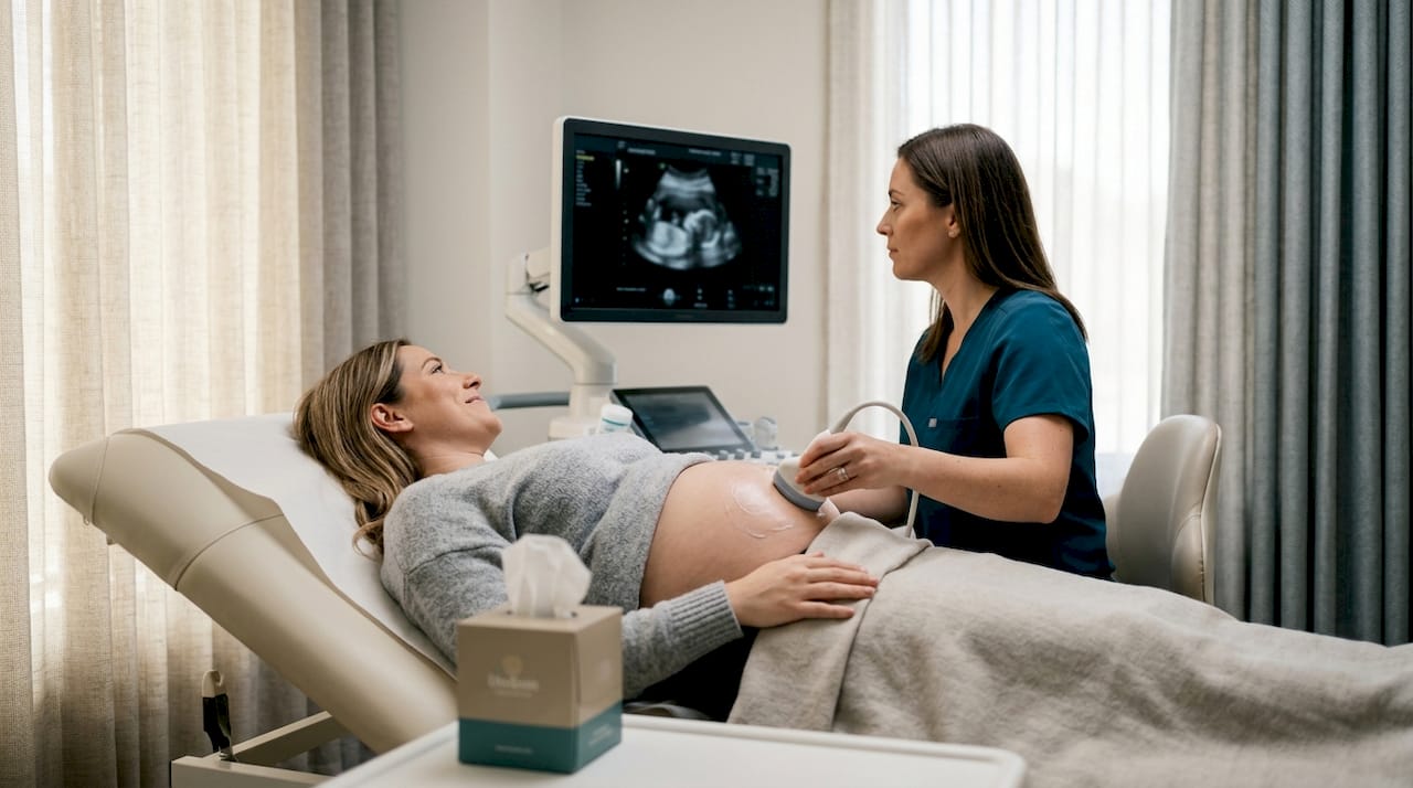
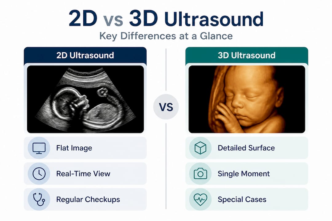
A 2D ultrasound produces flat, cross-sectional images of your baby in real time. Think of it as a slice through the body, showing internal structures like the heart, brain, kidneys, and spine in shades of grey. This is the image most people associate with a standard prenatal scan. It is the workhorse of obstetric imaging, capable of measuring growth, assessing fluid levels, checking fetal position, and evaluating organ development.
A 3D ultrasound, by contrast, reconstructs multiple 2D images into three-dimensional still images, providing detailed views of fetal surface features like the face, limbs, and body contours. This technology is also useful for detecting specific anomalies such as cleft lip or spinal defects that may be harder to see in 2D. The result is an image that looks like a photograph of your baby, showing recognisable facial features and surface details.
Understanding the 2D vs 3D vs 4D comparison is especially helpful because many families assume that more dimensions automatically means better medical information. That is not always the case.
| Feature | 2D ultrasound | 3D ultrasound |
|---|---|---|
| Image type | Flat, cross-sectional | Three-dimensional, surface-rendered |
| Primary use | Diagnostic, routine prenatal | Adjunct, elective, surface detail |
| Structures seen | Internal organs, blood flow | Surface features, facial contours |
| Covered by OHIP | Yes (routine) | No (elective) |
| Availability | Standard at all clinics | Specialised equipment required |
| Scan time | Shorter | Longer |
Key differences between 2D and 3D ultrasounds at a glance:
- 2D shows internal structures in real time, essential for measuring growth and detecting internal anomalies
- 3D creates still images of the baby’s outer surface, including face, hands, and feet
- 4D is simply 3D in motion, showing the baby moving in real time
- Ultrasound technology advancements have made 3D imaging more accessible, but its clinical role remains defined and limited
How ultrasounds are performed: technical requirements
After understanding the core image differences, let’s look at what goes on behind the scenes and what you should expect during your appointment.
A standard 2D ultrasound uses a single-plane transducer, a handheld probe that the sonographer (ultrasound technologist) moves across your abdomen or, earlier in pregnancy, inserts vaginally. The probe emits sound waves, receives the echoes, and converts them instantly into a 2D image on screen. It is a relatively straightforward process that requires skilled interpretation but not specialised hardware beyond the standard machine.
Mechanically, 3D ultrasound requires specialized transducers such as mechanical sweeping probes or matrix arrays, or sophisticated software, to acquire and process volumetric data from multiple 2D slices. The equipment is more expensive, and the operator must be trained not only to capture the images correctly but also to manipulate the volumetric data to produce a useful result.
| Requirement | 2D ultrasound | 3D ultrasound |
|---|---|---|
| Transducer type | Standard single-plane | Specialised volumetric or sweeping |
| Operator training | Standard sonography training | Advanced 3D acquisition skills |
| Scan duration | Typically 20 to 45 minutes | Often longer depending on purpose |
| Equipment cost | Lower | Higher |
| Image processing | Real-time | Post-processing required |
It is also worth noting that 3D is better for obese patients or situations with a poor 2D window, but this advantage comes with a trade-off: it requires higher skill, a longer scan time, and the quality of the 3D image depends entirely on the quality of the underlying 2D data. If the 2D acquisition is poor, the 3D rendering will be too.
Pro Tip: Before booking a 3D scan at any clinic, ask specifically whether their sonographers hold advanced training or certification in 3D acquisition. The quality of your images depends just as much on the operator as it does on the machine itself.
When you visit Valence Medical Imaging’s ultrasound equipment, you can see the kind of investment in technology that matters when it comes to producing reliable diagnostic images.
When and why each ultrasound is used in Ontario prenatal care
Now that you’ve seen how ultrasounds work, let’s connect those details to Ontario’s medical system and how families actually access these scans.
In prenatal care, 2D is standard, medically indicated, and covered by OHIP in Ontario for routine scans. A typical pregnancy includes at least one or two 2D scans, one in the first trimester to confirm the pregnancy and estimate gestational age, and an anatomy scan around 18 to 20 weeks to assess fetal development in detail. If there are concerns about growth, placenta position, amniotic fluid, or fetal wellbeing, additional scans may be ordered by your physician or midwife. All of these are 2D scans.
3D and 4D scans, by contrast, are elective, non-diagnostic, and private-pay. They are not considered part of routine prenatal care in Ontario. Boutique clinics across Southern Ontario offer these scans primarily for bonding and keepsake purposes. The cost is paid out of pocket and is typically not reimbursed through any provincial or private health plan.
Medical organisations such as ACOG, AIUM, and SOGC emphasise 2D for routine prenatal ultrasound, recommending 3D only as an adjunct for problem-solving rather than routine care, due to no proven added benefit and the potential for over-reliance on surface imaging.
“The best scan is the one your provider recommends for your baby’s health. Choosing an ultrasound type based on image aesthetics rather than clinical guidance can give families a false sense of security.”
Reading about prenatal ultrasound in Toronto is a good starting point if you want to understand what is routinely offered and covered in this province.
There are, however, legitimate clinical situations where 3D imaging adds value:
- Surface anomaly detection: When a 2D scan raises a concern about cleft lip, limb abnormalities, or neural tube defects, 3D imaging may provide more detail to support the clinical decision
- Poor 2D acoustic window: In some situations, such as maternal body habitus or unfavourable fetal position, a 3D approach may yield better surface information
- Emotional bonding: For families who have experienced pregnancy loss or anxiety, seeing a clear facial image of their baby can have genuine emotional value, though this must be understood as an add-on, not a replacement for standard care
- Specific fetal positions: When the fetus is positioned in a way that limits 2D views of particular structures, 3D reconstruction can sometimes fill the gap
Understanding the role of routine prenatal evaluations in Ontario’s healthcare system helps families set realistic expectations and avoid unnecessary costs.
Safety and emotional benefits of prenatal ultrasounds
Having covered medical necessity, let’s address your concerns about both the safety and the emotional experience these ultrasound technologies offer.
All types of prenatal ultrasound are safe, using non-ionizing sound waves rather than radiation. This means there is no known risk of harm to the baby or mother from the sound waves themselves. Both 2D and 3D ultrasounds use the same underlying technology; the difference is in how the data is captured and presented. However, Canadian guidelines recommend that elective 3D and 4D scans be performed for medical purposes only, not simply for entertainment or keepsake photography.
That said, the emotional benefits that families report from 3D imaging are real and not to be dismissed. Seeing your baby’s face clearly for the first time can be a powerful and memorable experience. For families navigating anxiety, previous losses, or simply wanting to feel more connected to their pregnancy, a clear 3D image can provide meaningful reassurance.
Here are the key benefits that both types of scans can offer, each in their appropriate context:
- Confirmation of healthy development: Both 2D and 3D scans reassure families that the pregnancy is progressing as expected
- Early anomaly detection: 2D remains the primary tool for identifying structural problems early in pregnancy
- Surface detail for specific concerns: 3D adds value when your provider wants a closer look at facial or limb structures
- Family connection: Sharing ultrasound images with partners, grandparents, and siblings creates a sense of early bonding with the new baby
- Emotional reassurance during high-risk pregnancies: Seeing a moving, developing baby can support mental health and reduce anxiety for parents who are carrying higher levels of stress
- Supporting provider decisions: Both types of imaging contribute to a fuller clinical picture when providers are managing complex pregnancies
Pro Tip: Always prioritise scans recommended by your midwife, obstetrician, or family physician. If you are considering an elective 3D scan, confirm that the facility follows Canadian professional guidelines and uses certified diagnostic imaging staff.
For a clear breakdown of ultrasound safety during pregnancy, you will find reliable, clinically grounded information that addresses the most common concerns expectant parents raise.
Our perspective: what families actually need to know about 2D vs 3D ultrasounds
With the facts in hand, here is what we have learned from more than 35 years of supporting families across Southern Ontario in making informed imaging choices.
The single most important thing we see families get wrong is assuming that a more visually impressive scan is a more medically useful one. A beautiful 3D image of your baby’s face is genuinely special. But it tells your doctor far less than a clear 2D view of the four chambers of the heart. Diagnostic accuracy is not measured in how lifelike an image looks.
We have also noticed that social media has shifted expectations significantly. Parents arrive having seen their friends’ vivid 3D scans and feel that anything less is somehow inadequate. That is understandable, but it is not medically sound. A standard 2D anatomy scan performed by a skilled sonographer on well-maintained equipment will always provide your provider with more actionable health information than a 3D keepsake session.
Another pattern we observe is that families sometimes book elective 3D scans without informing their healthcare provider. This can lead to confusion about results, particularly if the 3D image raises a concern that is then not communicated back to the clinical team. Always loop in your provider before and after any imaging, elective or otherwise.
Our genuine advice is this: trust your provider’s guidance on what scans you need, when, and why. If you choose to add an elective 3D scan for emotional reasons, do so through a reputable facility with trained staff. Do not let it replace the medically indicated scans your OHIP coverage provides.
Families across Southern Ontario who want to navigate their ultrasound options thoughtfully will find that the standard medical pathway, supported by a quality diagnostic imaging provider, is more than sufficient for monitoring a healthy pregnancy.
Where to get quality ultrasound services in Southern Ontario
As you finish your research and prepare for your own scan, here is where you can access top-quality services and guidance close to home.
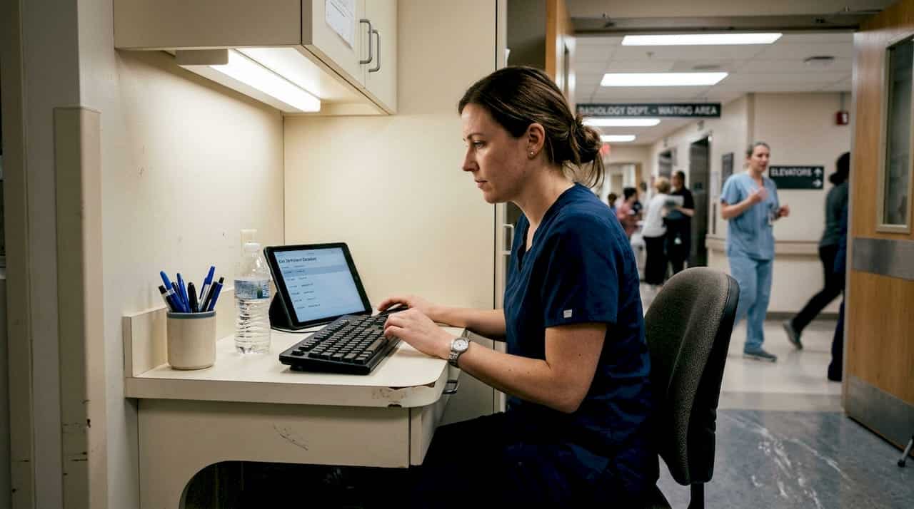
Valence Medical Imaging has been serving families across Southern Ontario for over 35 years, with seven clinic locations in Toronto, Scarborough, Brampton, Bramalea, Niagara Falls, and Whitby. Our Ontario ultrasound clinic locations are staffed by trained, experienced sonographers using high-end diagnostic equipment. We offer short wait times, fast report turnaround, and a patient-first experience that makes every appointment as comfortable and efficient as possible.
Whether your physician has referred you for a routine prenatal scan or you have questions about what imaging is appropriate for your stage of pregnancy, our team is ready to help. You can also explore a detailed guide on how ultrasound works to feel fully prepared before your appointment. Booking is simple and straightforward, and we welcome both patient-initiated and provider-referred appointments.
Frequently asked questions
Is 3D ultrasound covered by OHIP in Ontario?
No, 3D ultrasound is considered elective and is not covered by OHIP. Standard 2D prenatal scans are medically indicated and covered for routine care in Ontario.
Are 3D ultrasounds safe for my baby?
Yes, all prenatal ultrasound types are safe, using non-ionizing sound waves. However, Canadian guidelines recommend that elective 3D and 4D scans be performed for medical reasons rather than purely for entertainment.
Can 3D ultrasound detect problems that 2D cannot?
3D ultrasound reconstructs multiple 2D images to show surface features, making it more helpful for detecting specific anomalies like cleft lip or spinal defects. However, routine prenatal screening is still performed with 2D ultrasound as the primary tool.
Do I need a 3D ultrasound if my 2D scan was unclear?
In some situations, yes. 3D is better for poor 2D windows, such as cases involving maternal obesity or specific anomaly suspicion, though it requires a more skilled operator and a longer scan time. Your provider will advise whether a 3D scan is appropriate for your situation.
Recommended
- What is the difference between a 2D, 3D, and 4D ultrasound? – 2D vs. 3D vs. 4D Ultrasounds: Comparing Imaging Techniques – Valence Medical Imaging
- Exploring 3D and 4D Ultrasounds in Ontario: The Future of Imaging – Valence Medical Imaging
- The Essential Role of Regular Ultrasounds in Ontario Healthcare – Valence Medical Imaging
- Are 4D Ultrasounds Safe for Babies? – Bringing Baby to Life in Real-Time – Valence Medical Imaging
- Ultrasonido prenatal: guía para una experiencia emotiva