Ultrasound has quietly become one of the most relied-upon diagnostic tools in Southern Ontario, yet many patients still think of it only as a prenatal scan. The reality is far broader. Utilization has nearly doubled in hospitalised patients between 2007 and 2017, rising from 2.3% to 4.5%, and that growth continues in outpatient clinics across the province. From confirming a healthy pregnancy to screening for bone loss after menopause, ultrasound covers a remarkable range of health needs safely and efficiently. This article walks you through how ultrasound works, when it is used, and how to access timely services right here in Southern Ontario.
Table of Contents
- What makes ultrasound uniquely safe and effective
- Ultrasound in prenatal care: Setting standards for safety and detail
- Assessing bone quality: Quantitative ultrasound vs. traditional DXA
- Getting timely diagnostic ultrasound in Ontario: Practical patient advice
- Ultrasound’s evolving impact: What most patients and providers miss
- Connect with trusted ultrasound services in Southern Ontario
- Frequently asked questions
Key Takeaways
| Point | Details |
|---|---|
| Radiation-free imaging | Ultrasound offers real-time medical imaging without exposing patients to ionizing radiation. |
| Essential for prenatal care | Ultrasound is the primary tool for monitoring pregnancy, assessing fetal development, and confirming viability. |
| Early osteoporosis screening | Quantitative Ultrasound is an accessible first step for bone density assessment, but DXA follow-up is required for diagnosis. |
| Timely access advice | Booking with a prompt physician referral and checking wait times can help patients get scans quickly in Ontario. |
| Operator skill matters | The quality of your ultrasound results depends significantly on the experience of the technician performing your scan. |
What makes ultrasound uniquely safe and effective
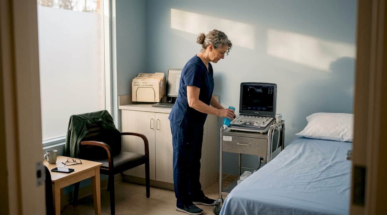
Most patients know that X-rays involve radiation, but not everyone realises that ultrasound involves none at all. Instead of radiation, ultrasound relies on high-frequency sound waves generated by a piezoelectric transducer, a small crystal inside the probe that converts electrical energy into sound. Those sound waves travel into body tissue, bounce back as echoes when they hit different structures, and are converted into real-time images on a screen. The whole process happens in milliseconds, which is why a sonographer can watch your heart beating or your baby moving in live footage.
Because there is no ionising radiation involved, ultrasound is considered safe across all age groups. It is the preferred imaging tool for pregnant patients, newborns, and children, and it can be repeated as often as clinically necessary without any cumulative risk. That is a meaningful advantage over CT scanning, which carries a radiation dose that physicians must weigh carefully before ordering.
Beyond safety, ultrasound is cost-effective and accessible. Equipment can be portable enough to fit at a bedside or in a remote clinic, and most scans produce results almost immediately. Sonographers can guide physicians in real time, which matters enormously in time-sensitive situations. The role of regular ultrasounds in preventive and ongoing care is increasingly recognised by Ontario clinicians.
Advantages of ultrasound at a glance:
- No ionising radiation, safe for all patients including pregnant women and children
- Real-time imaging allows dynamic assessment of movement and blood flow
- Portable units can reach community clinics, remote locations, and bedside in hospital
- Faster turnaround than MRI with no need for breath-holding sequences
- Lower cost compared to MRI or CT, making it highly accessible under OHIP
| Feature | Ultrasound | CT scan | MRI |
|---|---|---|---|
| Radiation exposure | None | Moderate to high | None |
| Real-time imaging | Yes | No | Limited |
| Soft tissue detail | Good | Moderate | Excellent |
| Cost | Low | Moderate | High |
| Wait time (typical Ontario) | Short | Moderate | Long |
| Safe in pregnancy | Yes | No | Conditional |
“Ultrasound is the only modality that gives us real-time, radiation-free images of soft tissues and blood flow simultaneously, making it indispensable for a wide range of clinical situations.” This reflects what sonographers and referring physicians across Ontario observe daily in practice.
Pro Tip: If your physician orders an ultrasound and you are uncertain why, ask whether it is to assess structure, blood flow, or both. Knowing which type of scan you are having, whether abdominopelvic, Doppler, or musculoskeletal, helps you prepare and ask the right questions.
Now that you see why ultrasound is so widely used and safe, let us explore its main roles across different types of health assessments in Ontario.
Ultrasound in prenatal care: Setting standards for safety and detail
Prenatal ultrasound is the application most Ontarians are familiar with, but there is a lot more clinical precision behind it than many expect. It is not simply about seeing the baby. Each scan serves a specific medical purpose tied to the stage of pregnancy, and following recommended timing improves the quality of information gathered.
Prenatal ultrasound is standard for confirming viability, estimating gestational age, detecting fetal anomalies, and measuring nuchal translucency, a fluid accumulation at the back of the neck that can indicate chromosomal conditions like Down syndrome. The World Health Organization recommends at least one ultrasound scan before 24 weeks of pregnancy. Ontario protocols generally follow a similar schedule, with many providers recommending scans at approximately 11 to 13 weeks and again at 18 to 20 weeks.
Recommended prenatal ultrasound schedule in Ontario:
- First trimester (11 to 13 weeks): Confirms viability and heartbeat, estimates gestational age, and measures nuchal translucency as part of the integrated prenatal screen.
- Second trimester anatomy scan (18 to 20 weeks): Examines fetal organ development, placental position, amniotic fluid levels, and growth parameters.
- Third trimester (if indicated): Assesses fetal growth, position, and placental function when specific concerns arise.
Understanding early pregnancy ultrasounds helps expecting parents know what to expect at each visit. Many patients feel reassured when they understand the difference between a scan that checks anatomy and one that monitors growth.
| Assessment | Ultrasound | MRI |
|---|---|---|
| Gestational age estimation | Excellent | Not routinely used |
| Fetal anatomy survey | Very good | Superior for brain and spine detail |
| Nuchal translucency measurement | Gold standard | Not applicable |
| Placental position | Excellent | Excellent |
| Radiation risk | None | None |
| Availability in Ontario | Wide | Limited, longer wait |
| Cost to patient (OHIP covered) | Yes (with referral) | Conditional |
MRI is reserved for specific situations where ultrasound findings are inconclusive, particularly for fetal brain or spinal cord assessment. For the vast majority of pregnancies, ultrasound provides everything clinicians need.
Expectant parents in Toronto and across Southern Ontario can find prenatal ultrasound in Toronto services with short wait times and experienced sonographers. If you are navigating your choices, reviewing ultrasound options for pregnancy can help clarify what is available and what your referral covers.
Pro Tip: Drink the recommended amount of water before your first trimester scan as instructed by your clinic. A full bladder lifts the uterus and provides a clearer acoustic window, particularly early in pregnancy before the uterus is large enough to be easily seen.
After understanding prenatal protocols, let us look at how ultrasound supports bone health and screens for osteoporosis risk.
Assessing bone quality: Quantitative ultrasound vs. traditional DXA
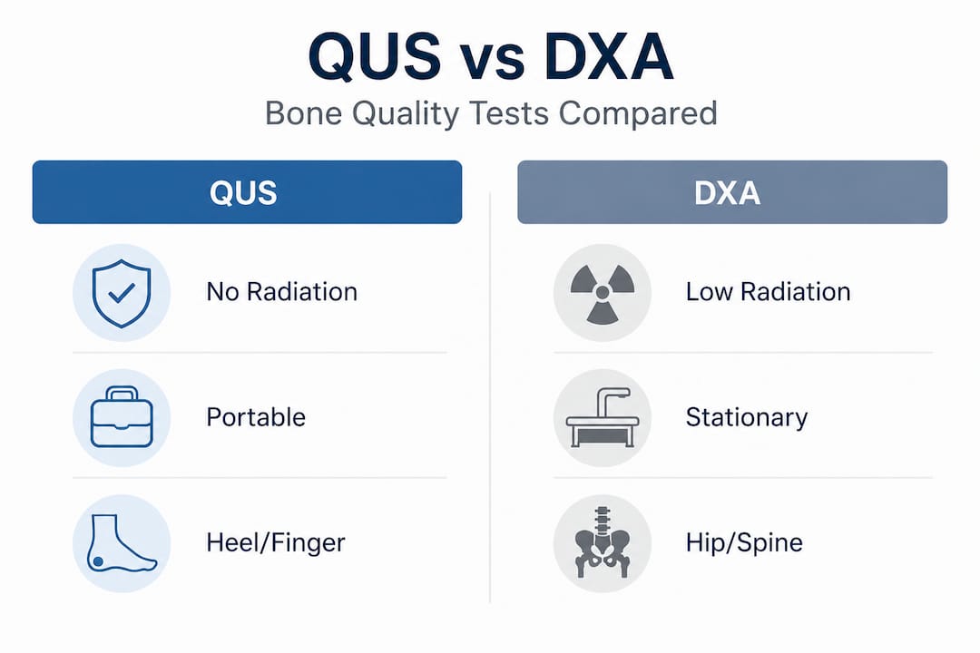
Most people associate bone density testing with a separate X-ray-based machine called DXA (dual-energy X-ray absorptiometry). DXA remains the gold standard for diagnosing osteoporosis and tracking treatment response over time. But there is a radiation-free alternative worth knowing about, particularly for initial community-level screening.
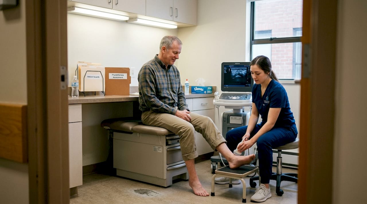
Quantitative ultrasound, or QUS, measures bone quality at peripheral sites such as the heel or finger bones. It does this by assessing two key parameters: broadband ultrasound attenuation (BUA), which measures how sound energy is absorbed by bone, and speed of sound (SOS), which measures how fast sound travels through bone. Together, these values provide an index of bone density and structure without any radiation exposure.
QUS is particularly useful for postmenopausal women who may need initial screening but do not yet have access to a DXA machine, or who are being monitored in a community setting. Its portability makes it practical for family health teams and community clinics.
Who may benefit from QUS screening:
- Postmenopausal women aged 50 and over
- Patients with a family history of osteoporosis
- Individuals who have experienced a low-impact fracture
- Those who cannot access DXA promptly
| Feature | QUS | DXA |
|---|---|---|
| Radiation exposure | None | Very low |
| Portability | High | Low |
| Site measured | Heel, phalanx | Hip, spine |
| Osteoporosis diagnosis | Screening only | Gold standard |
| Fracture risk prediction | Moderate | High |
| Cost | Lower | Moderate |
| Ontario availability | Community clinics | Specialised centres |
Key limitation: QUS does not account for bone size or volume the way DXA does, which means a positive QUS screen should always be followed up with DXA for a formal diagnosis. It is a starting point, not a finishing line.
Patients in Brampton and surrounding areas can explore bone density test options available locally. For those who want a broader overview of what the process involves, learning about bone scan procedures provides a useful foundation before your appointment.
Bone quality assessment is just one example. Let us examine practical tips for patients who want timely access to diagnostic ultrasound across Ontario.
Getting timely diagnostic ultrasound in Ontario: Practical patient advice
Access to diagnostic imaging in Ontario can vary considerably depending on where you live, the urgency of your referral, and whether you are seeking care in a hospital or an independent clinic. The good news is that with the right preparation, you can reduce delays and arrive at your appointment fully ready.
Step-by-step guide to booking your ultrasound:
- Obtain a referral: Most OHIP-covered ultrasounds require a physician, nurse practitioner, or midwife referral. Physician referrals are the most direct route, and referring providers can often flag urgency to speed up access.
- Check local wait times: Ontario Health publishes wait time data that can help you understand typical delays in your region and identify facilities with shorter queues.
- Choose the right setting: Independent imaging clinics often have shorter wait times than hospital radiology departments for non-urgent referrals. Comparing hospital vs clinic ultrasounds helps you make an informed decision.
- Confirm preparation requirements: Different scans have different prep instructions. Abdominal ultrasounds typically require fasting for four to six hours and a full bladder. Pelvic ultrasounds may require a full bladder only. Vascular Doppler studies usually have no prep requirements.
- Bring your paperwork: Carry your requisition, health card, and any previous imaging reports. Relevant prior studies give the interpreting radiologist important context.
Common reasons for delays and how to avoid them:
- Incomplete or missing requisition: Ask your physician to confirm the referral was sent before your appointment.
- Insufficient bladder preparation: Follow the clinic’s hydration instructions precisely.
- Arriving without prior imaging: Collect relevant reports and bring them to your appointment.
- Booking in a high-volume hospital department for a routine scan: Consider an independent clinic for non-urgent studies.
It is also worth knowing the limitations of ultrasound so you are not surprised if additional imaging is recommended. Some anomalies require MRI for clearer detail, particularly in the brain and spine, and ultrasound image quality is affected by patient body habitus, bowel gas, and operator experience. Understanding the ultrasound referral process in Ontario removes a lot of confusion before you even make your first call.
Pro Tip: If you are unsure which common Ontario ultrasound procedures apply to your situation, call the clinic ahead of your appointment. Sonographers and booking staff can confirm your preparation requirements and clarify any questions about what the scan will assess.
With these practical tools in hand, let us reflect on the wider impact and lessons learned about ultrasound’s evolving role in Ontario.
Ultrasound’s evolving impact: What most patients and providers miss
Here is something worth saying plainly: the technology itself matters far less than the skill of the person performing the scan. Ultrasound is operator-dependent in ways that CT and MRI are not. A CT machine captures a fixed dataset that any radiologist can review. An ultrasound scan is an active, real-time process where the sonographer’s technique, probe placement, and clinical judgment determine what gets captured and what gets missed.
This is particularly true for complex assessments like nuchal translucency measurement in early pregnancy, where a difference of a fraction of a millimetre can shift a risk calculation significantly. It is also true for vascular Doppler studies, where the angle of insonation (the direction sound waves hit a blood vessel) directly affects the accuracy of blood flow measurements. Patients asking whether a clinic has experienced, credentialed sonographers are asking exactly the right question.
There is a parallel issue with QUS in community screening. Portable QUS devices are genuinely valuable for making bone health screening more accessible across Southern Ontario, particularly in smaller communities or practices that cannot accommodate a full DXA suite. But patients and clinicians alike sometimes treat a reassuring QUS result as a clear bill of bone health. It is not. QUS screens; it does not diagnose. Any elevated risk identified by QUS should be followed by DXA and a proper assessment with a healthcare provider who understands fracture risk tools and clinical history.
Another overlooked reality is the importance of context. The same ultrasound image can lead to very different clinical decisions depending on the patient’s history, symptoms, and prior imaging. A small fibroid identified incidentally is managed very differently in a 30-year-old versus a 58-year-old. Radiology reports are most useful when paired with a conversation between the patient and their referring physician, rather than being read in isolation. Ultrasound’s role in Toronto’s healthcare ecosystem is growing precisely because clinicians are learning to integrate it earlier and more strategically into care pathways.
The takeaway is this: access to good imaging equipment is necessary, but not sufficient. The right scan, performed by an experienced sonographer, read by a knowledgeable radiologist, and interpreted in clinical context by an informed physician gives you the best possible outcome. Choose your imaging provider thoughtfully.
Connect with trusted ultrasound services in Southern Ontario
At Valence Medical Imaging, we have spent over 35 years supporting patients and physicians across Southern Ontario with reliable, timely diagnostic imaging. Whether you are expecting a baby, managing bone health concerns, or following up on a new symptom, our seven clinic locations in Toronto, Scarborough, Brampton, Bramalea, Niagara Falls, and Whitby are equipped to help. If you are new to diagnostic imaging, our resources on how ultrasounds work provide a straightforward starting point. To understand when your doctor might refer you, our guide on reasons for ultrasound exams covers the most common clinical situations clearly. You can also review our overview of Ontario ultrasound procedures to see the full range of services available. We welcome same-day bookings for select services and offer fast report turnaround so your care team can act on results promptly.
Frequently asked questions
Is ultrasound covered by OHIP in Ontario?
Most medically necessary ultrasounds are covered by OHIP for Ontario residents who have a valid health card and a physician or authorised healthcare provider referral. Some elective or non-referred scans, such as keepsake pregnancy ultrasounds, are not covered.
Are ultrasounds safe during pregnancy?
Yes. Ultrasounds are safe for pregnant women and their babies because they use sound waves rather than ionising radiation, and no confirmed harmful effects have been identified at diagnostic levels.
What preparation is needed for an abdominal ultrasound?
You will typically be asked to fast for four to six hours and arrive with a full bladder, as preparation significantly improves image quality. Always follow the specific instructions provided by your clinic, as requirements can vary by scan type.
Does QUS replace DXA for osteoporosis diagnosis?
No. QUS complements but does not replace DXA for diagnosing osteoporosis. QUS is a useful radiation-free screening tool, but DXA remains the gold standard for formal diagnosis and treatment monitoring.
What limits the accuracy of ultrasound imaging?
Image quality is operator-dependent and can be reduced by patient body habitus, bowel gas, bone structures, and air, which block sound wave transmission. Some clinical scenarios, such as detailed fetal brain assessment, may require MRI to supplement ultrasound findings.
Recommended
- The Essential Role of Regular Ultrasounds in Ontario Healthcare – Valence Medical Imaging
- Ultrasound’s Crucial Role in Toronto’s Healthcare Revolution – Valence Medical Imaging
- 5 Must-Know Ultrasound Procedures in Ontario Clinics – Valence Medical Imaging
- Cutting-Edge Ultrasound Therapies Transforming Toronto Healthcare – Valence Medical Imaging