X-rays and Fluoroscopy are two popularly used techniques for diagnosing diseases and illnesses. These imaging-based diagnostic techniques help the doctor get a clear image of what’s happening within the body. These procedures create a film or a set of images that help the doctor see the bones and the body’s internal structure. But, it is important to understand how these two techniques are different from one another.
What is an X-ray?
An X-ray is an image of the internal body structure that has been captured on an X-ray film. It uses radiation that has been projected directly onto the patient’s target site. A sensor is placed against the target site that helps produce the X-ray image. This image is static. Since it is purely based on the density of the bone, a certain amount of X-ray radiation manages to pass through the bone. Thus, the X-ray images are considered to show outdated information and don’t exhibit the current condition of the bones.
What are the uses of X-rays?
X-ray images are mainly used to check the static internal structures and identify the current condition. For instance, they are popularly used to determine trauma to bones, in case of an injury, such as fractures and hairline cracks. Multiple X-rays are taken and kept for record in cases of degenerative bone diseases, such as rheumatoid arthritis. This helps in understanding the degeneration rate and comparing the patient’s existing condition.
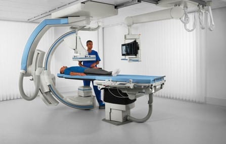
What is Fluoroscopy?
Fluoroscopy is an advanced diagnostic technique that is used to derive real-time images. In this technique, multiple X-ray beams are projected on the target site of the body. These X-ray beams are continuous. The images obtained are directly projected onto the computer screen with the help of advanced technology. Since multiple images are being created within a fraction of a second, it seems like multiple images are being created. This helps the doctor to get a real-time diagnosis and creates multiple images.
Why is Fluoroscopy used?
Due to the differences in the image production and quality of the image, doctors prefer using Fluoroscopy over X-rays for various techniques and diagnostic needs. Here are some of the most popular cases where doctors prefer using Fluoroscopy over X-rays-
- Fluoroscopy is actively used during the insertion of catheters. These small tubes must be inserted into the blood vessels and can be needed for different purposes. Since the blood vessels are thin and can have loops, bifurcations, constrictions, etc., it is important to view them in real-time. This helps in preventing ruptures and excessive bleeding. Fluoroscopy helps view the blood vessels in real-time, thus reducing complications. Moreover, it also helps in inserting stents and works great in reducing complications.
- Fluoroscopy is a great help in performing angiograms. They help assist doctors in a great way in viewing the real-time internal structure of the body. This technique helps in determining if there is anything wrong. Other procedures were Fluoroscopy for gastrointestinal diseases and orthopedic surgical procedures.
What is the difference between X-ray and Fluoroscopy?
There is a major difference between X-rays and Fluoroscopy, especially in image production. While X-rays are static and still images, Fluoroscopy produces multiple real-time images. Thus, the uses for Fluoroscopy are more in the medical industry. Here are two of the major differences between X-rays and Fluoroscopy-
- X-ray images are captured on the sensor with the help of a clicker. During this process, as soon as the button is clicked for image capturing, a sharp beam of X-ray photons is projected on the screen after passing through the target site. This process is a single-take; there is no scope for correction. Depending on the type of X-ray imaging used, the image is produced digitally or on a film. If the image is distorted or the positioning is incorrect, the entire process has to be repeated.
Compared to this, during Fluoroscopy, a continuous beam of X-ray photons is released on the target site, producing multiple images. As the sensor moves, the target site and angle shift accordingly.
- The patient is exposed to a single beam of X-ray during an X-ray, while during a fluoroscopy, the patient is projected to multiple beams. This directly increases the amount of risk the patient has to face due to continuous radiation exposure. The patient may have to face serious complications if proper preventive measures are not taken. Thus, repeated use of Fluoroscopy is omitted for diagnosis.
Conclusion
X-ray images are static and produce old images. On the other hand, Fluoroscopy offers better diagnosis and helps in creating multiple real-time moving images. X-ray images are comparatively inexpensive compared to Fluoroscopy. At the same time, Fluoroscopy has major side effects as the amount of radiation exposure is comparatively much higher as compared to normal X-rays. Irrespective of the side effects of Fluoroscopy, they are a preferable and more reliable method of diagnosis.