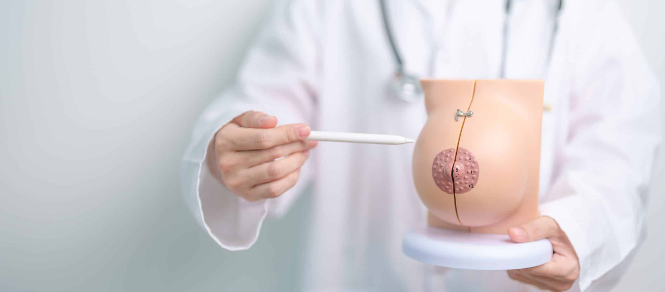
For women in Toronto, Brampton, Whitby, and Niagara Falls, and indeed across the world, breast imaging is an essential health procedure for the early detection and treatment of breast cancer. Valence Medical Imaging offers a range of advanced breast imaging techniques, including digital mammograms, which are pivotal in breast cancer screening programs. A clear understanding of breast anatomy is crucial to comprehend how these imaging techniques, particularly mammography, work and their role in detecting breast abnormalities.
The Basics of Breast Anatomy:
The female breast is composed of several types of tissues, mainly glandular, fibrous, and fatty tissues. Glandular tissue includes the lobules, which produce milk, and ducts, the tiny tubes that carry milk toward the nipple. Surrounding these are fibrous tissues and ligaments providing structural support, while fatty tissue encompasses these structures, giving the breast its size and shape.
Breast Imaging Techniques and Their Relation to Anatomy:
- Digital Mammogram: This is the most common form of breast imaging and involves taking X-ray pictures of the breast. It is designed to identify abnormalities in the breast tissue that cannot be felt during a physical exam. The digital aspect allows for better contrast resolution than traditional film, making the detailed anatomy of the breast more visible.
- Diagnostic Mammogram: When an initial screening mammogram detects an abnormality, or if there are signs of breast cancer, a diagnostic mammogram is performed. It is a more detailed X-ray of the breast, often involving additional views to closely examine the areas of concern.
- Breast Tomosynthesis: Also known as 3D mammography, breast tomosynthesis takes multiple X-ray pictures of each breast from different angles, providing a more complete picture of the breast tissue. This method can be particularly useful for women with dense breasts.
- Breast Ultrasound: Ultrasound uses sound waves to produce images of structures deep within the body. Breast ultrasound can complement mammography by providing additional information about a lump or abnormality detected on a mammogram.
- Breast Biopsy: If a suspicious area is identified on an imaging test, a biopsy may be necessary. This involves removing a sample of breast tissue to be examined under a microscope for signs of cancer.
Breast Density and Its Importance:
Breast density is a term used to describe the proportion of the different types of breast tissue visible on a mammogram. Dense breasts have higher amounts of glandular and fibrous tissue and less fatty tissue. Since dense tissue and tumors both appear white on a mammogram, it can be more difficult to detect cancer in women with dense breasts, which is where breast tomosynthesis can be especially beneficial.
The Role of BI-RADS Classification:
The Breast Imaging-Reporting and Data System (BI-RADS) classification is used by radiologists to categorize mammography findings. The BI-RADS score ranges from 0 to 6, with each number representing a different level of concern for cancer, and assists in determining the need for additional testing or biopsy.
Radiation Exposure:
While mammography exposes the breast to a small amount of radiation, the risk of harm is considered very low compared to the substantial benefits of early cancer detection. Valence Medical Imaging employs state-of-the-art technology to minimize radiation exposure while ensuring high-quality images.
Breast anatomy plays a significant role in how mammography and other imaging modalities are utilized for breast cancer screening and diagnosis. Valence Medical Imaging in the Greater Toronto Area is committed to offering women the most advanced breast imaging services, including digital mammograms, diagnostic mammograms, breast tomosynthesis, breast ultrasounds, and breast biopsies. With a deep understanding of breast anatomy and the latest technology, radiologists at Valence Medical Imaging can provide accurate assessments of breast health, ensuring that any signs of cancer are detected and addressed as early as possible. Remember, regular breast cancer screening is a vital step in protecting your health. Contact Valence Medical Imaging to schedule your appointment and for more information on the services available to you.


