Since the olden days, radiography has been a popular mode of diagnosis and checking the internal structures of the human body. They greatly help the doctors understand what is happening within the body and confirm their diagnosis. It also helps them identify if any other concerns might have been missed during the preliminary diagnosis. The X-ray principle is based on the density of the bones. This means, in reality, that the current x-ray shows the condition of the bone that was before a certain period.
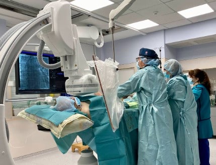
It can be said that an X-ray doesn’t showcase a real-time image. An advanced step of traditional X-rays is fluoroscopy. This is an advanced technique in which the doctors take a continuous X-ray which helps in viewing the internal structures of the body in real-time. Other than accurate diagnosis, fluoroscopy is also popularly being used to help surgeons during surgery and provide them with better results. Before understanding the benefits of fluoroscopy, let us first understand what it exactly is.
What is fluoroscopy?
To understand in a simple and common term, fluoroscopy can be titled a real-time moving x-ray. It helps the doctors to identify the exact condition of the patient and the disease that is present currently. Since the information is not outdated as it happens in a traditional x-ray, the doctor is in a better position to make an accurate diagnosis and devise a better treatment plan.
During an x-ray imaging, a single x-ray beam is passed through the patient via the target site. When this hits the x-ray film, an image is produced. This image is based on the density of the bone and is a static image. In reality, it can only cover and create images as per a particular percentage of bone density. Thus, in a way, it can be said that the image is actually outdated. On the other hand, during fluoroscopy, multiple X-ray beams are targeted on the patient’s target site. The images produced are directly projected on the computer screen to create a real-time moving image.
Why is fluoroscopy done?
The X-ray images are majorly used for checking out the diseases of the bones and have a limit when it comes to checking out the organs. On the other hand, fluoroscopy offers a better view of the internal and external organs of the body, along with the bones. Thus, it helps in an accurate and wider diagnosis. The doctors can easily view bones, muscles, joints, tendons, ligaments, and body systems. Spinal procedures are the most common reasons why doctors order fluoroscopy diagnostic tests. For spinal fluoroscopy tests, the doctor injects the dye directly into the spine via the spinal taps. This is also known as lumbar punctures. Or, they can also be directly injected into the spinal canals.
What are the benefits of fluoroscopy for spinal injections?
Fluoroscopy is a specialized diagnostic tool used to evaluate the meninges. During this process, a special dye is inserted within the spinal cord, that is a contrast material. This process is also known as myelography. After the insertion of the dye, fluoroscopy is done, where multiple real-time images can be seen on the computer screen. This helps the doctor view the nerve endings better. It is a popularly used diagnostic tool, especially in the cases of patients with packmarkers, who cannot undergo an MRI.
Another popular use of fluoroscopy within the spine is for guided orthopedic surgeries. Fluoroscopy helps the doctor understand the placement and movement of instruments better. It is commonly used in cases of trauma or spinal surgery.
Risks of fluoroscopy
Most popular medical procedures also have their own set of risks and benefits. The common reason for fluoroscopy risks is the excessive amount of radiation the patient is exposed to. Due to this, the patient may experience many side effects, including nerve damage and disabilities. If a female is pregnant, there can be permanent damage to the child’s health, including physical and mental deformities. Before agreeing to a fluoroscopy procedure, every female must get a pregnancy test done to confirm if she is pregnant. It must be completely avoided, in case she is.
The other most common risk factor for fluoroscopy is cancer. The radiation emitted during this process can greatly affect cell reproduction and control thus, can cause cancer. This type of cancer is known as radiation cancer. Other common complications of this diagnostic procedure include cross-infection due to needle contamination, excessive bleeding, etc.
Conclusion
Fluoroscopy is an advanced diagnostic technique with multiple X-ray beams passing through the patient’s body. This helps create a continuous image that can be broadcasted on a computer screen. There are many benefits of fluoroscopy, including better diagnosis and the ability to view the body’s internal structure. At the same time, it also has several risks associated with it. However, it can be said that the benefits of fluoroscopy outweigh its cons.


