Mammography remains one of the most prevalent methods for breast cancer screening, particularly in cities like Toronto, Brampton, Whitby, and Niagara Falls. A mammogram aims to detect early signs of breast cancer, ideally before symptoms even begin. However, like all medical screenings, mammograms aren’t infallible. Misinterpretations can sometimes occur, leading to what’s known as false positives and false negatives.
Understanding Mammography
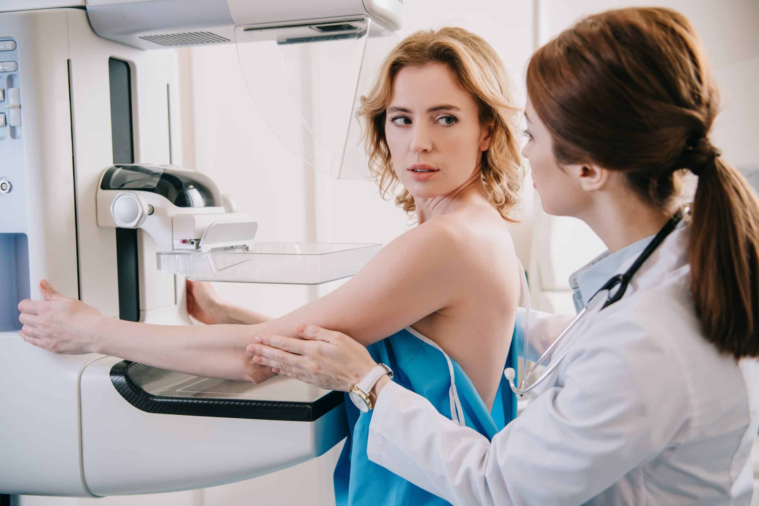
Before we delve into the intricacies of false results, let’s gain an understanding of the mammography process itself.
Digital Mammogram: Unlike traditional film mammograms, digital mammograms capture and store the image electronically. This makes it easier for radiologists at centers like Valence Medical Imaging in Toronto to zoom in, manipulate the images, and ensure clarity, which can be crucial for those with dense breasts.
Breast Tomosynthesis: Often termed as 3D mammography, breast tomosynthesis offers multiple images or slices of the breast from different angles. This increases the chances of spotting a potential cancerous lesion, especially in women with dense breast tissue.
What are False Positives and Negatives?
False Positive: This occurs when a mammogram indicates that breast cancer might be present when it is not. This can lead to unnecessary stress, further testing like a diagnostic mammogram, breast ultrasound, or even a breast biopsy.
False Negative: On the other hand, a false negative is when a mammogram appears normal even though breast cancer is present.
Factors Leading to False Positives and Negatives
Dense Breasts: Women with dense breasts have more connective and glandular tissue compared to fatty tissue. This can sometimes obscure or mimic tumors, leading to a false positive. On the flip side, it can also hide tumors, leading to a false negative.
Breast Imaging Experience: The skill level of the radiologist can also play a role. BI-RADS Classification, or Breast Imaging-Reporting and Data System, provides radiologists with standardized criteria for categorizing mammographic findings. However, if the radiologist is unfamiliar with certain nuances, it could result in misinterpretation.
Radiation Exposure: While mammograms expose women to a small amount of radiation, it’s typically not enough to increase the risk of breast cancer. However, multiple screenings or other types of diagnostic tools can increase the total radiation exposure, albeit the risk remains minimal.
Mitigating the Risk
Breast Ultrasound: This can be used as an adjunct to mammography, especially in women with dense breasts. It can help distinguish between fluid-filled cysts (which are typically non-cancerous) and solid masses.
Breast Biopsy: If an abnormality is detected, a biopsy can provide a definitive answer. It involves removing a small amount of tissue to be examined under a microscope.
Regular Screening: Despite the possibility of false results, regular breast cancer screening remains crucial. Catching cancer early can make a significant difference in outcomes.
At Valence Medical Imaging in Toronto, and our branches in Brampton, Whitby, and Niagara Falls, our commitment is to provide the highest level of care and accuracy in breast imaging. Being informed about the potential for false positives and negatives, understanding the factors that contribute to them, and knowing how they are managed, empowers patients to make informed decisions about their health. Remember, while mammography isn’t perfect, it remains an invaluable tool in the fight against breast cancer.