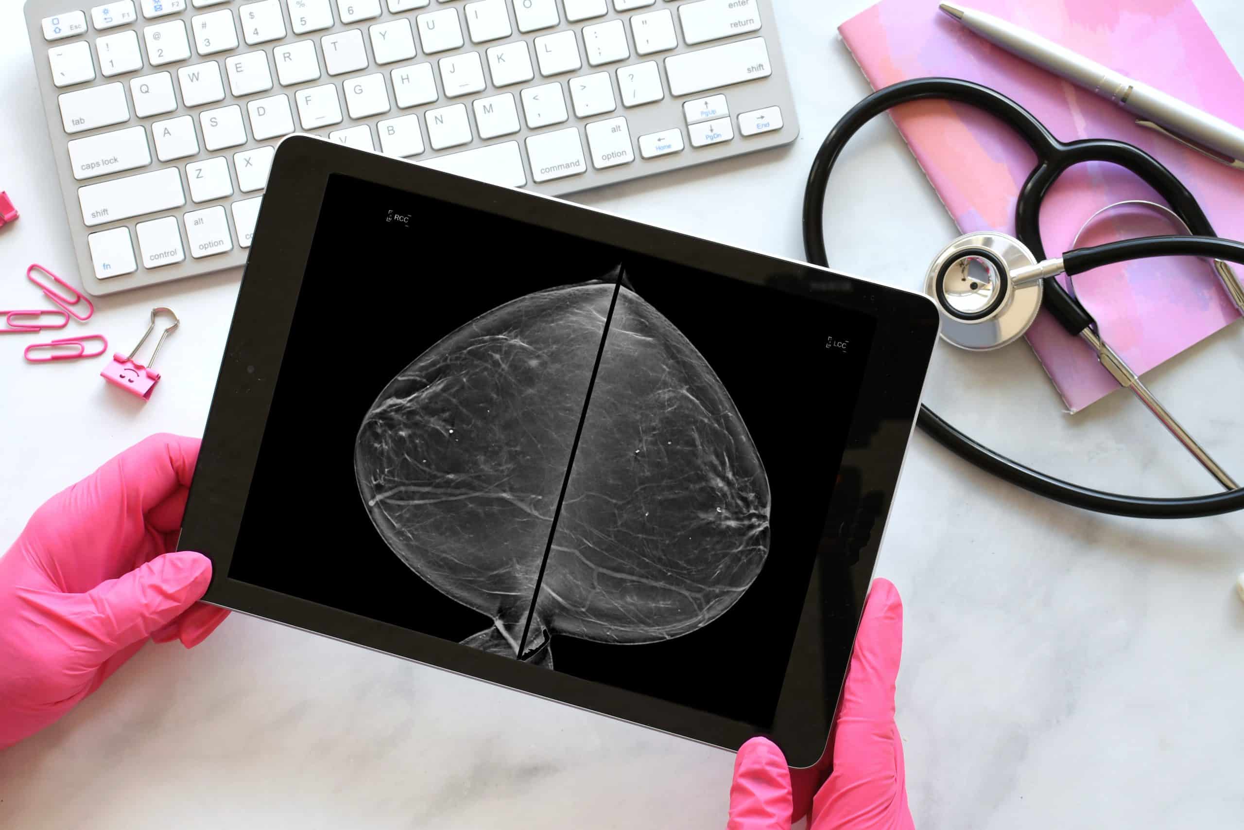
As we delve deeper into the world of medical imaging, specifically within the realm of breast imaging, the tools and techniques at our disposal continually evolve. These innovations pave the way for more accurate and less invasive diagnosis, supporting early detection and enhancing patient outcomes. Located in Toronto and serving surrounding areas including Brampton, Whitby, and Niagara Falls, Valence Medical Imaging is at the forefront of these advancements. Let’s explore the cutting-edge methods and how they are revolutionizing breast cancer screening.
Digital Mammogram: The Modern Approach
Traditionally, mammograms relied on film-based techniques. However, the advent of digital mammography offers clearer images and quicker results. The ability to digitally manipulate the image to adjust contrast or zoom in on suspicious areas means that radiologists can make more accurate assessments, reducing the number of unnecessary callbacks.
BI-RADS Classification: A Standardized Reporting System
The Breast Imaging Reporting and Data System (BI-RADS) classification ensures consistency in mammogram reporting. This standardized system categorizes breast imaging findings on a scale from 0 (incomplete) to 6 (proven malignancy), enabling healthcare professionals to communicate with clarity and take subsequent steps with precision.
Reducing Radiation Exposure
One of the concerns with mammograms has always been radiation exposure. But with the latest technology, radiation doses have been significantly reduced without compromising image quality. This makes breast cancer screening safer and encourages regular check-ups.
Understanding Dense Breasts
Dense breast tissue can sometimes obscure cancer on a mammogram. However, advancements in imaging techniques like breast tomosynthesis and breast ultrasound have provided improved imaging solutions for women with dense breasts, making early detection more feasible.
Diagnostic Mammogram: A Deeper Look
Should a screening mammogram detect an anomaly, a diagnostic mammogram takes a closer, more detailed look. This high-resolution imaging focuses specifically on the area of concern, ensuring that any abnormalities are clearly identified and assessed.
Breast Tomosynthesis: A 3D Perspective
Also known as 3D mammography, breast tomosynthesis captures multiple images of the breast from different angles, creating a three-dimensional image. This technique offers a clearer view of overlapping tissues and can pinpoint abnormalities with enhanced accuracy compared to traditional 2D imaging.
Breast Biopsy Imaging
When a suspicious area is identified, a breast biopsy might be recommended. Imaging-guided biopsies, often using mammography or ultrasound, ensure precise targeting of the lesion, making the procedure less invasive and more accurate.
The Role of Breast Ultrasound
Breast ultrasound uses sound waves to produce images of the breast’s internal structures. Especially valuable for women with dense breasts, it can distinguish between solid masses (potentially cancerous) and cysts (fluid-filled and typically non-cancerous). The procedure complements mammography and provides additional valuable diagnostic information.
The world of breast imaging is in the midst of rapid and profound transformation. The primary goal remains the early detection of breast cancer and reducing the morbidity associated with delayed diagnosis. With advancements like digital mammograms, BI-RADS classification, and the incorporation of 3D imaging techniques, Valence Medical Imaging in Toronto stands poised to deliver top-notch breast imaging services to the communities of Brampton, Whitby, and Niagara Falls, ensuring that every woman has access to the best in breast cancer screening technology.


