In an era marked by rapid advancements in medical technology, it’s imperative for patients and healthcare professionals alike to stay informed about the most effective diagnostic tools available. Mammography, a critical tool in breast cancer detection, has evolved significantly over the years. Today, both digital and film mammography options are available, each with its own set of advantages and considerations. For the residents of Toronto, Brampton, Whitby, and Niagara Falls, understanding the distinctions between these two methods is essential in making informed choices about their healthcare. This article delves into the specifics of both digital and film mammography, providing clear insights to help you navigate your screening options in the age of modern medical imaging.
The Evolution of Mammography
Film Mammography: The original mammography technique, film mammography or “film-screen mammography”, uses low-dose X-rays to capture images of the breast on film. These films are then developed in a darkroom.
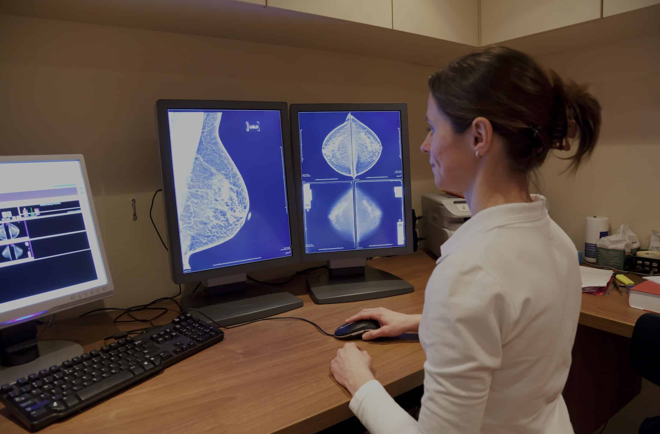
Digital Mammography: Also known as full-field digital mammography (FFDM), digital mammography uses electronic detectors to capture the X-ray images and display them on a computer. This digital format allows for immediate viewing and manipulation of images.
Advantages of Digital Mammography
1. Image Manipulation: Radiologists can adjust the brightness, contrast, and zoom of a digital mammogram, making it easier to spot abnormalities.
2. Better for Dense Breasts: Studies have shown that digital mammography can be more accurate in women with dense breasts. The ability to adjust the image can help radiologists see through dense tissue.
3. Storage and Transfer: Digital images are stored electronically, making them easier to share with other health professionals or facilities.
4. Less Radiation Exposure: While all mammography techniques use low doses of radiation, some studies suggest that digital methods may use up to 25% less radiation than film mammograms.
The Advent of Breast Tomosynthesis
Building upon digital mammography, breast tomosynthesis or “3D mammography” captures multiple X-ray images from different angles. These images are then reconstructed into a three-dimensional model, allowing for more detailed examination.
Benefits:
- Improved detection rates.
- Reduction in false positives, which can reduce the need for follow-up testing or breast biopsy.
- Enhanced clarity, especially in dense breasts.
When is Additional Testing Necessary?
Sometimes a mammogram—whether film or digital—indicates the need for further testing.
- Breast Ultrasound: Often used for women with dense breasts, this imaging technique uses sound waves to capture images of the breast tissue.
- Diagnostic Mammogram: If an abnormality is found in the screening mammogram, a diagnostic mammogram focuses on the area of concern using various angles.
- Breast Biopsy: If a suspicious area is detected, a small sample of tissue is removed for examination.
Understanding the BI-RADS Classification
The Breast Imaging Reporting and Data System (BI-RADS) classification helps radiologists communicate the findings of a mammogram. This system ranks the results on a scale from 0 (incomplete) to 6 (known biopsy-proven malignancy), guiding the next steps in the patient’s care.
Making an Informed Choice
With advancements in breast imaging, patients have more options than ever. Whether opting for a digital mammogram, breast tomosynthesis, or other imaging techniques, the primary goal remains early and accurate detection of breast cancer.
For residents in Toronto, Brampton, Whitby, and Niagara Falls, Valence Medical Imaging remains at the forefront of these innovations, ensuring that each patient receives the best care tailored to their individual needs.
Remember, the best mammogram is the one that gets done. Whether you’re due for your routine breast cancer screening or have specific concerns, don’t hesitate to consult with us to help guide you through the process.


