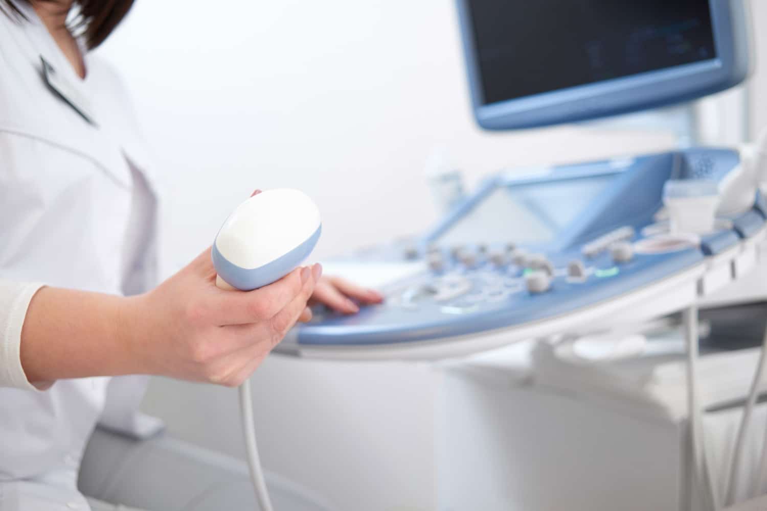
Ultrasound imaging or sonography is an advanced innovation in the field of medicine. Typically they are best known for capturing images of the growing fetus. However, this imaging test is beneficial in vascular medicine as a primary diagnostic tool that creates live images of blood flow in your vessels.
But why should you do the vascular ultrasound? Vascular ultrasound is sued to detect the flow of blood throughout the vessels. It will help the physicians learn about what is happening to the arteries in your heart and the blood vessels in your legs.
You can properly diagnose your vascular problems and create an appropriate treatment plan. Keep reading to discover more about the significance and types of vascular ultrasound!
Vascular Ultrasound – An Overview
A vascular ultrasound exam is non-invasive and painless. Vascular Ultrasound uses sound waves to produce images of blood vessels present within the circulatory system of your body. This procedure is generally used to examine the artery or vein in any part of your body, including the blood vessels in the abdominal region, neck, and lower and upper extremities.
Diagnosing or ruling out any vascular diseases, including deep vein thrombosis, gallbladder disease, and more, is useful. This procedure uses no harmful ionizing radiation, so there will be no harmful effects.
Why Should You Do Vascular Ultrasound?
As a vital component of our circulatory system, a heart beats 60 to 100 times in a minute. Each heartbeat pumps oxygenated blood into your body, which travels through the collective system of arteries and blood vessels. Arteries deliver nutrients and oxygen to all the organs and tissues and then return the blood to the heart and lungs for reoxygenation. Together, they comprise the vascular or circulatory system.
The circulation cycle is an ongoing process to ensure that oxygenated blood gets delivered to all body parts while deoxygenated blood returns to the heart. When any damage or infection to this system is left undiagnosed, it will lead to life-threatening conditions. Vascular ultrasounds are designed to examine the parts of this circulatory/vascular system, including veins, arteries, and capillaries.
More specifically, it will be suggested to detect the blow flow in the arteries in your neck that deliver blood to the brain or blood flow to the transplanted organ and to detect the presence, severy, and location of the narrowed or blockage of blood flow in the arteries. Your physician might require you to undergo this vascular ultrasound if you have experienced the following symptoms.
- Nausea
- Chest pain
- Sweating
- Troubled or shortness of breath
- Appearance or pale or bluish shade in your skin
- Difficulty in swallowing
- Restriction in mobility
- Feeling tired or weak for a prolonged period
- Severe pain that steadily progresses throughout the body.
How to Prepare for Vascular Ultrasound?
There are no strict or specific preparation rules for the vascular ultrasound examination. Most physicals will recommend wearing loose-fitting and comfortable clothing during the procedure. It is because you will be instructed to remove the clothing in the target area. If the process is suggested to detect the vessels in the abdominal region, you must be fasting.
What Happens During Vascular Ultrasound?
You are instructed to lie down on the exam table in a face-up position. The ultrasound process will begin with a technical applying a cooling gel to the target area. During the vascular ultrasound procedure, a small probe coated with gels will be placed in contact with the skin of a specific area. The probe will send high=freinquen waves which have no radiation. This device, the transducer, is used to convert one form of energy to another to have smooth and secure contact.
The sound waves used in the vascular sound will be transmitted throughout the tissues of the target site that need to be examined. They will reflect the blood flow inside the blood vessel. When the waves bounce back, the transducer collects them and converts them into a computerized image. The entire process will take around 30 to 90 minutes to complete. Even though it’s a painless process, you may experience a little discomfort when applying pressure on your arms or legs with the transducer.
Why Do I Need a Vascular Ultrasound Scan?
Vascular imaging is commonly used in the diagnostic procedure to assess the location and severity of any ailments. In general, vascular ultrasound examinations are performed to
- Monitor blood flow to particular organs and tissues
- Identify the location of blockages, narrowing (vascular stenosis), or abnormalities in the blood vessels.
- Detect the speed of blood flow
- Identify the formation of blood clots
- Determine the enlargement of the blood vessels
- Assess the development of varicose veins
- Determine whether an angioplasty is required
- Analyze the success rate of a surgical procedure
What are the Types of Vascular Ultrasound?
An ultrasound screening is a non-invasive procedure. Depending on the individual’s symptoms, the physician will recommend a specific type of vascular ultrasound. Here we have listed out the types of ultrasound.
- Arterial Doppler Ultrasound
Arterial Doppler Ultrasound is a type of imaging test recommended when the person shows poor arterial or venous circulation symptoms. It may affect the neck, legs, or arms. Decreased blood flow occurs due to blood clots, blood vessel injury, or arterial blockage.
An arterial Doppler ultrasound will measure the pressure of the targeted vessels and detect the speed of blood flow. It is beneficial in identifying peripheral artery disease and other venous-related implications.
- Carotid Ultrasound
Carotid ultrasound is the screening procedure performed on one side of your neck, where the carotid arteries are situated. This ultrasound assesses the blood in these arteries and diagnoses the presence of any blockage or arterial plaque that will eventually result in atherosclerosis when left noticed.
- Renal Artery Ultrasound
The renal artery ultrasound procedure is used to evaluate the arteries that play a vital role in the oxygenation and nourishment of kidneys. It will detect the narrowing or blockage in these arteries, contributing to hypertension and renal dysfunction.
What to Expect After Vascular Ultrasound?
Once the vascular ultrasound procedure is completed, you can drive yourself to your home and carry on regular activities on the same day as it is non-invasive. You will also receive the results from the technical immediately. Once the results are received, your physician will promptly discuss them with you to create a personalized treatment approach to heal your conditions.
Wrapping Up!
Learning about the status of your vascular health plays a key part in your comprehensive heart care. Vascular ultrasounds are performed to examine how well your blood is flowing throughout your body. This procedure is quick and painless, posing no health risks. Ultrasound technology is a reliable way to prevent the onset of vascular diseases from leading a healthy, active life. Are you seeking the perfect partner for your cardiovascular health? We are here to help! At Valence Medical Imaging, we offer various types of vascular ultrasound to observe your blood flow through the vessels. With complete diagnostic services, we ensure to accurately diagnose and treat our patients as efficiently and quickly as possible.


