Since 1896, diagnosis and treatment planning has improved drastically. The introduction of fluoroscopy by Enrico Salvioni into the field of medicine has helped greatly in improving the identification of dental diseases and disorders and understanding the real-time need for treatment protocol.
However, fluoroscopy was limited in dental procedures due to the high radiation. As image intensification was introduced in the later years, the need for higher radiation was reduced. Ever since then, its implications and demand have increased.
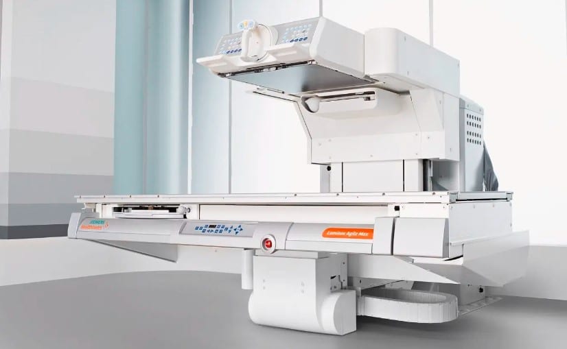
Fluoroscopy in Diagnostic Procedures
The process of fluoroscopy has changed the approach to diagnostic procedures, which has helped improve treatment planning.
It has helped improve the patient’s care and enhanced the prognosis and treatment output. Fluoroscopy is a diagnostic technique that works quite similarly to an X-ray. There are many differences, which makes it more accurate.
In X-rays, radiations are passed through the body part, which helps in producing radiographic imaging. These images are static and help doctors identify the condition of the bone. However, in cases of bone degeneration, about 30% of changes cannot be determined due to the difference in mineral composition.
This makes fluoroscopy a more accurate diagnostic technique. It is far more reliable as it shows real-time changes.
Unlike X-rays, where only static images are produced, multiple images are produced during fluoroscopy and can be directly viewed on the screen. Thus, making it convenient to be used during procedures. Let us understand the implications of fluoroscopy in detail –
How is Fluoroscopy Used in Diagnostic Procedures?
There are several different ways in which fluoroscopy can be used during diagnostic procedures. Here are four main uses of fluoroscopy-
- Barium Swallow Test
A barium swallow test is a special diagnostic test used to identify the complications and issues in the esophagus, stomach, and a part of the small intestine. In this test, the patient is made to swallow a special barium dye.
This test is commonly used to diagnose esophageal webs, stomach ulcers, tumors, GERD (gastro-oesophageal reflux disorder), etc. The barium helps mark and spot pathological changes, which helps identify diseases.
During the barium swallow test, the doctor uses the method of fluoroscopy to obtain real-time images of what’s happening inside the body. This helps ensure the dye is in the right position and helps identify the pathological changes.
- Swallow Study
A swallow study is a test in which the doctor examines your ability to swallow food items of different textures, ranging from solid to liquid. In most studies, the patient is first made to swallow a barium dye that helps mark the esophagus and other parts.
During the swallow test, the doctor uses fluoroscopy to get images of the patient’s insides while swallowing food items. With the help of these images, the doctor closely examines the trachea, bronchi, esophagus, and pharynx. The doctor may also ask you to change your body posture while swallowing, such as head tilting, lifting your chin, etc.
- Cardiac Procedures
Cardiologists actively use fluoroscopy to help with faster and more real-time diagnosis. This can be life-saving during several sensitive operations and procedures.
Cardiac Catheterization
One of the most common cardiac procedures where fluoroscopy is indicated is during cardiac catheterization. In many patients, due to several reasons, such as obesity, high cholesterol, structural changes, etc., the cardiac artery constricts. This causes the cardiac blood output during pumping to reduce, reducing oxygen availability in the body.
In severe cases, this can also be life-threatening. Thus, the patients are advised for cardiac catheterization.
In this process, the doctor inserts a small catheter, a tube inside the cardiac artery. Once it reaches the right place, it is inserted and attached.
Fluoroscopy ensures that the catheter passes through the cardiac artery accurately. It also helps in confirming the site for insertion and ensures that there are no complications.
Angiography and Angioplasty
In many patients, a constriction or obstruction is seen in the passage of the cardiac artery. It is important to eliminate this obstruction to ensure smooth blood flow throughout the body. To identify the blockage, angiography is performed.
During this technique, the cardiologist passes a small tube within the body to identify the blockage. Also, fluoroscopy captures real-time moving images that help with the diagnosis. Fluoroscopy is also used on angioplasty, where the cardiologist removes these obstructions.
- Spinal Surgery
The use of fluoroscopy in spinal surgeries is highly recommended. The spine is one the most sensitive parts of the human body, as most nerve endings pass through. During spinal surgeries and administering injections, the doctor must ensure extreme precautions.
While several procedures, such as vertical body augmentation, spinal disk surgeries, insertion of the pedicle screws, etc., surgeons prefer using the fluoroscopy diagnostic technique. This helps them easily navigate and helps in reducing the risks associated with the surgeries.
Fluoroscopy is also used while the administration of spinal injections as it helps in identifying the exact location of the site of injection. It further helps to protect all vital organs in the surroundings.
Risks of Fluoroscopy
One of the most common potential risks associated with fluoroscopy is radiation exposure. The high amount of radiation used during fluoroscopy may cause damage to the underlying tissues and cells. Some cells may also burn due to the high amount of radiation.
Repeated exposure to these radiations may increase the chances of radiation cancer. In the case of pregnancy, these harmful radiations may negatively affect the growth of the fetus. It may even lead to miscarriage or premature delivery of the baby, risking its life.
How to Mitigate These Risks and Ensure Patient Safety?
Due to the high risks associated with fluoroscopy, it is not the first diagnostic technique used. Most doctors recommend other diagnostic techniques, such as X-rays, which are less invasive unless fluoroscopy is highly indicated.
It is important to take extensive measures while doing fluoroscopy. Here are some of the common steps doctors take to ensure patient safety and limit their radiation exposure
- The patients are made to wear a lead apron that helps in protecting them from the scattered radiation.
- A thyroid collar is recommended to prevent the risk of thyroid cancer.
- For less threatening procedures, the amount of exposure is limited and reduced to the bare minimum to ensure safety from radiation.
Conclusion
Fluoroscopy is a commonly used diagnostic technique in sensitive procedures, such as barium tests, swallow tests, cardiac surgeries, and spinal surgeries. This technique helps produce a series of real-time images that can be directly displayed on the screen.
Since multiple images are captured in a short interval, it seems a video is being captured and thus helps provide a sense of direction. Fluoroscopy has improved diagnosis and treatment planning and made it even more accurate. However, the doctors limit the radiation risk and take the necessary precautions to ensure patient care.